. 2019 May 7;19(1):424.
doi: 10.1186/s12885-019-5658-5.

Establishment of a rat ovarian peritoneal metastasis model to study pressurized intraperitoneal aerosol chemotherapy (PIPAC)

Affiliations

Establishment of a rat ovarian peritoneal metastasis model to study pressurized intraperitoneal aerosol chemotherapy (PIPAC)

Leen Van de Sande et al. BMC Cancer. .

Abstract

Background: pressurized intraperitoneal aerosol chemotherapy (PIPAC), with or without electrostatic precipitation (ePIPAC), was recently introduced in the treatment of peritoneal metastases (PM) from ovarian cancer (OC). Preliminary clinical data are promising, but several methodological issues as well the anticancer efficacy of PIPAC remain unaddressed. Here, we propose a rat ePIPAC model that allows to study these issues in a clinically relevant, reproducible, and high throughput model.

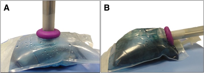
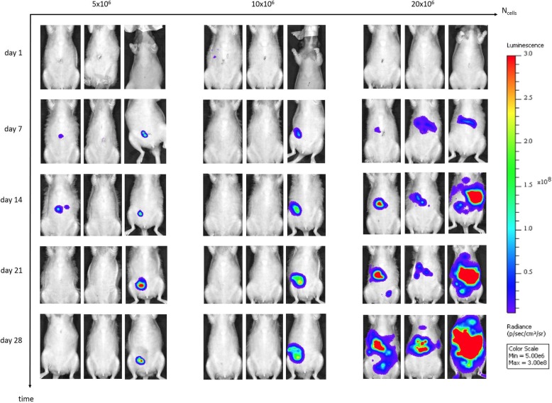
Methods: laparoscopy and PIPAC were established in healthy Wistar rats. Aerosol properties were measured using laser diffraction spectrometry based granulometric analyses. Electrostatic precipitation was accomplished using a commercially available generator (Ultravision™). A xenograft model of ovarian PM was created in athymic rats using intraperitoneal (IP) injection of SKOV-3 luciferase positive cells. Tumor growth was monitored weekly by in vivo bioluminescence imaging.

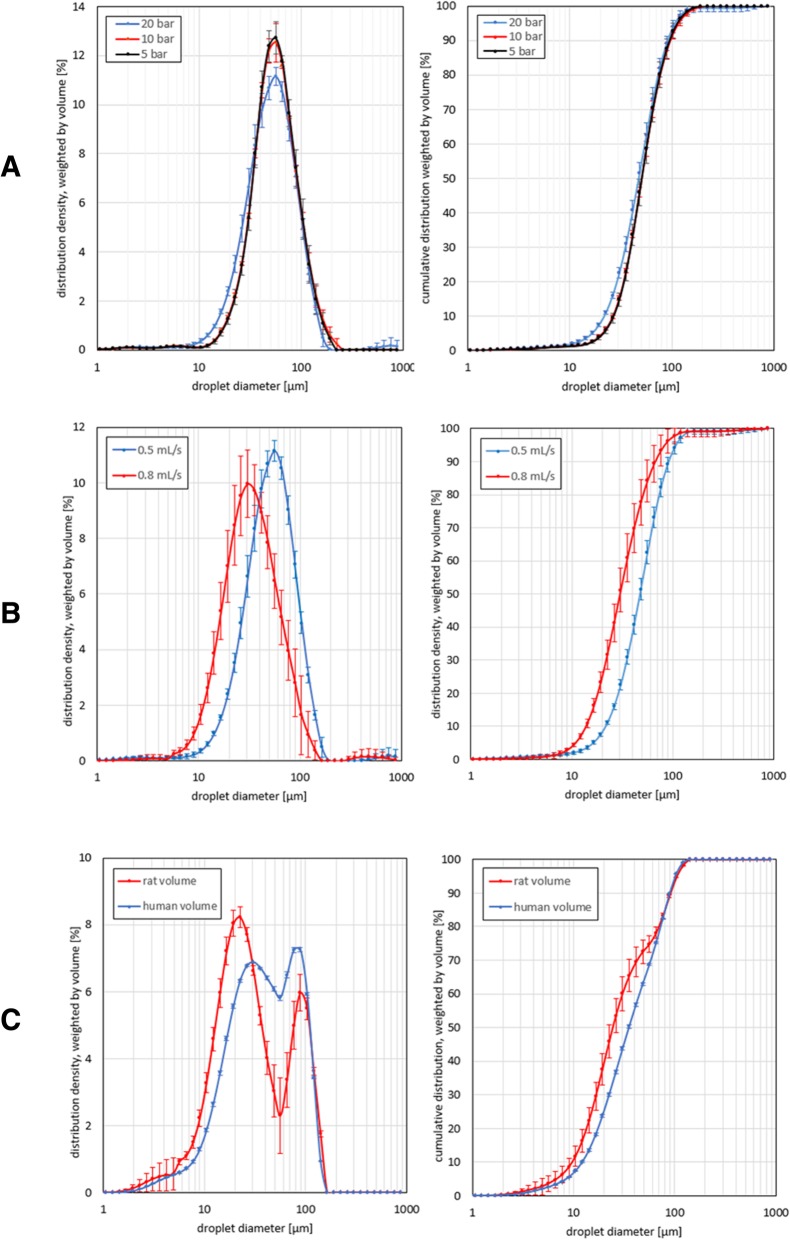
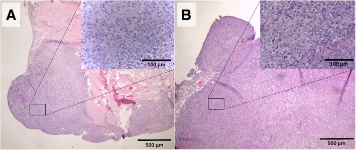
Results: PIPAC and electrostatic precipitation were well tolerated using a capnoperitoneum of 8 mmHg. All rats survived the (e)PIPAC procedure and no gas or aerosol leakage was observed over the entire procedure. With an injection pressure of 20 bar, granulometry showed a mean droplet diameter (D(v,0.5)) of 47 μm with a flow rate of 0.5 mL/s, and a significantly lower diameter (30 μm) when a flow rate of 0.8 mL/s was used. Experiments using IP injection of SKOV-3 luciferase positive cells showed that after IP injection of 20 × 106 cells, miliary PM was observed in all animals. PIPAC was feasible and well supported in these tumor bearing animals.

Conclusions: we propose a reproducible and efficient rodent model to study PIPAC and ePIPAC in OC xenografts with widespread PM. This model allows to characterize and optimize pharmacokinetic and biophysical parameters, and to evaluate the anti-cancer efficacy of (e)PIPAC treatment.

Keywords: Intraperitoneal drug delivery; Laparoscopic surgery; Ovarian cancer; PIPAC; Peritoneal metastases; Rat xenograft; ePIPAC.


Conflict of interest statement

Ethics approval

The study protocol was approved by the Animal Ethical Committee of the Faculty of Medicine, Ghent University, Belgium (ECD 17–50 and ECD 18–30). The human cell lines did not require ethics approval from our institution as they were purchased from legal commercial product.

Consent for publication

Not applicable.

Competing interests

The authors declare that they have no competing interests.

Publisher’s Note

Springer Nature remains neutral with regard to jurisdictional claims in published maps and institutional affiliations.

Figures

Fig. 1
Fig. 1
A ePIPAC setup in a healthy Wistar Hannover rat. 12 mm balloon trocar with nebulizer and closed aerosol waste system (a), 5 mm balloon trocar with laparoscope and CO2 insufflator (b), Ionwand (c), electrical conductor between return electrode (underneath metal plate) and generator unit (d). B Intra-abdominal view of the Ionwand in a healthy Wistar Hannover rat
Fig. 2
Fig. 2
PSD curves of nebulized saline showing the distribution density (left curves) and cumulative distribution (right curves). Mean droplet diameters were measured (n = 3 for each confirmation) in a range of 0.5 to 900 μm. The error bars show one time the standard deviation. a Volume-weighted PSD curves showing the influence of maximal upstream injection pressure on D(v,0.5). 20 mL of saline was nebulized in open space at a fixed flow rate of 0.5 mL/s and a maximal upstream injection pressure of 20 bar (blue graph), 10 bar (red graph) or 5 bar (black graph). b Volume-weighted PSD curves illustrating the influence of flow rate on D(v,0.5). 20 mL of saline was nebulized in open space at a fixed maximal upstream injection pressure of 20 bar and a flow rate of 0.5 mL/s (blue graph) or 0.8 mL/s (red graph). c Volume-weighted PSD curves demonstrating the influence of the volume of the peritoneal cavity on D(v,0.5). To simulate a nebulization in a rat’s abdominal cavity, 20 mL of saline was nebulized in a plastic box (V = 100 mL) with a flow rate of 0.8 mL/s and a maximal upstream injection pressure of 20 bar (red graph). 200 mL of saline was nebulized in a plastic box (V = 5 L) with a flow rate of 0.5 mL/s and a maximal upstream injection pressure of 20 bar (blue graph) as a comparison with the nebulization in human. The plastic boxes were bilaterally pierced to transvers laser light and a third perforation was made at the top of the boxes to insert the nebulizer
Fig. 3
Fig. 3
Distribution pattern of 20 mL undiluted royal blue ink. The injection parameters were set on a flow rate of 0.8 mL/s, a maximal upstream injection pressure of 20 bar and an intracavitary pressure of 8 mmHg. a Blue ink distribution was limited to the bottom of the EVA bag when the nebulizer was perpendicularly secured. b Complete staining of the EVA bag was observed when the nebulizer was placed in a tilted position
Fig. 4
Fig. 4
In vivo BLI of athymic nude rats inoculated with 5 × 106 (group 1), 10 × 106 (group 2) or 20 × 106 (group 3) SKOV-3 Luc IP2 cells (n = 3 in each group). In the first and second group no diffuse PM was observed. One rat demonstrated tumor clearance, two rats had only one tumor nodule and in the other specimens no tumor growth occurred. In the third group diffuse PM was observed with a tumor induction rate of 100%
Fig. 5
Fig. 5
Tumor nodules (white arrows) in athymic nude rats inoculated intraperitoneally with 20 × 106 SKOV-3 Luc IP2 cells. a Laparoscopic image of the right upper abdomen. b Post-mortem view of the upper abdomen. c Intestines and mesentery
Fig. 6
Fig. 6
H&E stainings of peritoneal implants at the parietal peritoneum (a) and mesentery (b), 40x magnification. In the right upper corner, a close up of the H&E staining is shown, 400x magnification. The typical morphology of cancer cells can be detected

References

    1. Al Rawahi T, Lopes AD, Bristow RE, Bryant A, Elattar A, Chattopadhyay S, Galaal K. Surgical cytoreduction for recurrent epithelial ovarian cancer. Cochrane Database Syst Rev. 2013;(2):CD008765. 10.1002/14651858.CD008765.pub3. - PMC - PubMed
    1. Oseledchyk A, Zivanovic O. Intraoperative Hyperthermic intraperitoneal chemotherapy in patients with advanced ovarian Cancer. Oncology (Williston Park, NY) 2015;29:695–701. - PubMed
    1. Klaver YLB, Simkens LHJ, Lemmens VEPP, Koopman M, Teerenstra S, Bleichrodt RP, de Hingh IHJT, Punt CJA. Outcomes of colorectal cancer patients with peritoneal carcinomatosis treated with chemotherapy with and without targeted therapy. Eur J Surg Oncol. 2012;38:617–623. doi: 10.1016/j.ejso.2012.03.008. - DOI - PubMed
    1. Thomassen I, van Gestel YR, van Ramshorst B, Luyer MD, Bosscha K, Nienhuijs SW, Lemmens VE, de Hingh IH. Peritoneal carcinomatosis of gastric origin: a population-based study on incidence, survival and risk factors. Int J Cancer. 2014;134:622–628. doi: 10.1002/ijc.28373. - DOI - PubMed
    1. Thomassen I, Lemmens VEPP, Nienhuijs SW, Luyer MD, Klaver YL, de Hingh IHJT. Incidence, prognosis, and possible treatment strategies of peritoneal carcinomatosis of pancreatic origin: a population-based study. Pancreas. 2013;42:72–75. doi: 10.1097/MPA.0b013e31825abf8c. - DOI - PubMed

MeSH terms