. 2019 May 21;39(5):BSR20190225.
doi: 10.1042/BSR20190225. Print 2019 May 31.

Oxidative and endoplasmic reticulum stresses are involved in palmitic acid-induced H9c2 cell apoptosis

Affiliations

Oxidative and endoplasmic reticulum stresses are involved in palmitic acid-induced H9c2 cell apoptosis

Lei Yang et al. Biosci Rep. .

Abstract

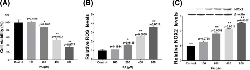
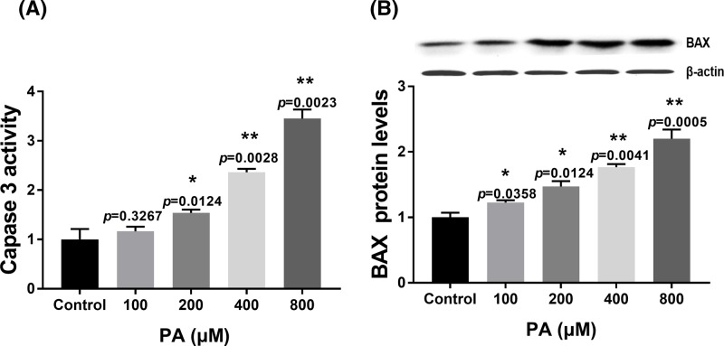
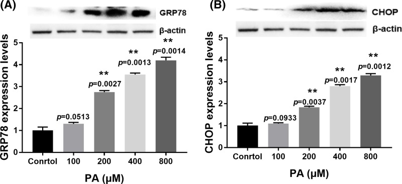
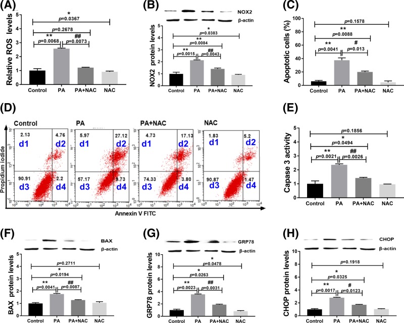
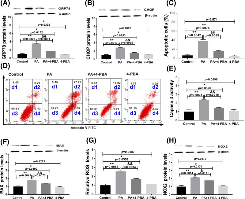
Palmitic acid (PA) is the most common saturated long-chain fatty acid that causes damage to heart muscle cells. However, the molecular mechanism of PA toxicity in myocardial cells is not fully understood. In the present study, we explored the effects of PA on proliferation and apoptosis of H9c2 cardiomyocytes, and uncovered the signaling pathways involved in PA toxicity. Our study revealed induction of both oxidative and endoplasmic reticulum (ER) stresses and exacerbation of apoptosis in PA-treated H9c2 cells. Inhibition of oxidative stress by N-acetylcysteine (NAC) reduced apoptosis and decreased ER stress in PA-treated H9c2 cells. Moreover, inhibition of ER stress by 4-phenyl butyric acid decreased apoptosis and attenuated oxidative stress. In summary, the present study demonstrated that oxidative stress coordinates with ER stress to play important roles in PA-induced H9c2 cell apoptosis.

Keywords: H9c2 cells; apoptosis; endoplasmic reticulum stress; oxidative stress; palmitic acid.


Conflict of interest statement

The authors declare that there are no competing interests associated with the manuscript.

Figures

Figure 1
Figure 1
Effect of PA on proliferation and oxidative stress of H9c2 cells (A) Cells were treated with various concentrations (100–800 µM) of PA for 24 h and then processed for cell viability analysis. (B) Cells were treated with various concentrations (100–800 µM) of PA for 24 h and then processed for the ROS assay. (C) Cells were treated with various concentrations (100–800 µM) of PA for 24 h and then analyzed for NOX2 expression. NOX2 expression was normalized to β-actin levels. Data are presented as the mean ± S.E.M. of three independent experiments, *P<0.05, **P<0.01 versus the control group.
Figure 2
Figure 2
Effect of PA on apoptosis-related protein expression in H9c2 cells (A) Caspase 3 activity in H9c2 cells after treatment with various doses of PA (100–800 µM) for 24 h. (B) Relative BAX expression in H9c2 cells after treatment with various doses of PA (100–800 µM) for 24 h. BAX expression was normalized to β-actin levels. Data are presented as the mean ± S.E.M. of three independent experiments, *P<0.05, **P<0.01 versus the control group.
Figure 3
Figure 3
Effect of PA on ER stress-related protein expression in H9c2 cells (A) Relative expression of GRP78 in H9c2 cells after treatment with various doses of PA (100–800 µM) for 24 h. (B) Relative expression of CHOP after treatment with various doses of PA (100–800 µM) for 24 h. Protein expression was normalized to β-actin levels. Data are presented as the mean ± S.E.M. of three independent experiments, 0.05<**P<0.01 versus the control group.
Figure 4
Figure 4
Effect of NAC on PA-induced apoptosis and ER stress of H9c2 cells at 24 h (A) ROS levels in H9c2 cells after treatments; (B) NOX2 expression levels in H9c2 cells after treatments. (C,D) Apoptosis analysis by flow cytometry. d1: necrotic cells; d2: late apoptotic cells; d3: live cells; d4: early apoptotic cells. (E) Caspase 3 activity in H9c2 cells after treatments; (F) BAX expression levels in H9c2 cells after treatments. (G) GRP78 expression levels in H9c2 cells after treatments; (H) CHOP expression levels in H9c2 cells after treatments. Protein expression levels were normalized to β-actin levels. PA: 400 µM palmitic acid; NAC: 2 mM N-acetylcysteine. Data are presented as the mean ± S.E.M. of three independent experiments, *P<0.05, **P<0.01 versus the control group and #P<0.05, ##P<0.01 represent PA+NAC treated group versus the PA-treated group.
Figure 5
Figure 5
Effect of 4-PBA on PA-induced apoptosis and oxidative stress of H9c2 cells at 24 h (A) GRP78 expression levels in H9c2 cells after treatments. (B) CHOP expression levels in H9c2 cells after treatments. (C,D) Apoptosis analysis by flow cytometry. d1: necrotic cells; d2: late apoptotic cells; d3: live cells; d4: early apoptotic cells. (E) Caspase 3 activity in H9c2 cells after treatments. (F) BAX expression levels in H9c2 cells after treatments. (G) ROS levels in H9c2 cells after treatments. (H) NOX2 expression levels in H9c2 cells after treatments. Protein expression levels were normalized to β-actin levels. PA: 400 µM palmitic acid; 4-PBA: 500 nM 4-PBA. Data are presented as the mean ± S.E.M. of three independent experiments, *P<0.05, **P<0.01 versus the control group and &P<0.05, &&P<0.01 represent PA+4-PBA treated group versus the PA-treated group.

References

    1. Zou L., Li X., Wu N., Jia P., Liu C. and Jia D. (2017) Palmitate induces myocardial lipotoxic injury via the endoplasmic reticulum stress-mediated apoptosis pathway. Mol. Med. Rep. 16, 6934 10.3892/mmr.2017.7404 - DOI - PubMed
    1. Utz W., Engeli S., Haufe S., Kast P., Hermsdorf M., Wiesner S.. et al. (2011) Myocardial steatosis, cardiac remodelling and fitness in insulin-sensitive and insulin-resistant obese women. Heart 97, 1585–1589 10.1136/hrt.2011.224451 - DOI - PubMed
    1. Haffar T., Akoumi A. and Bousette N. (2016) Lipotoxic palmitate impairs the rate of β-oxidation and citric acid cycle flux in rat neonatal cardiomyocytes. Cell. Physiol. Biochem. 40, 969 10.1159/000453154 - DOI - PubMed
    1. Leroy C., Tricot S., Lacour B. and Grynberg A. (2008) Protective effect of eicosapentaenoic acid on palmitate-induced apoptosis in neonatal cardiomyocytes. Biochim. Biophys. Acta 1781, 685–693 10.1016/j.bbalip.2008.07.009 - DOI - PubMed
    1. Park M., Sabetski A., Chan Y.K., Turdi S. and Sweeney G. (2015) Palmitate induces ER stress and autophagy in H9c2 cells: implications for apoptosis and adiponectin resistance. J. Cell Physiol. 230, 630–639 10.1002/jcp.24781 - DOI - PubMed

Publication types