. 2019 Jul 15;79(14):3662-3675.
doi: 10.1158/0008-5472.CAN-18-3464. Epub 2019 May 7.

Preexisting Commensal Dysbiosis Is a Host-Intrinsic Regulator of Tissue Inflammation and Tumor Cell Dissemination in Hormone Receptor-Positive Breast Cancer

Affiliations

Preexisting Commensal Dysbiosis Is a Host-Intrinsic Regulator of Tissue Inflammation and Tumor Cell Dissemination in Hormone Receptor-Positive Breast Cancer

Claire Buchta Rosean et al. Cancer Res. .

Abstract

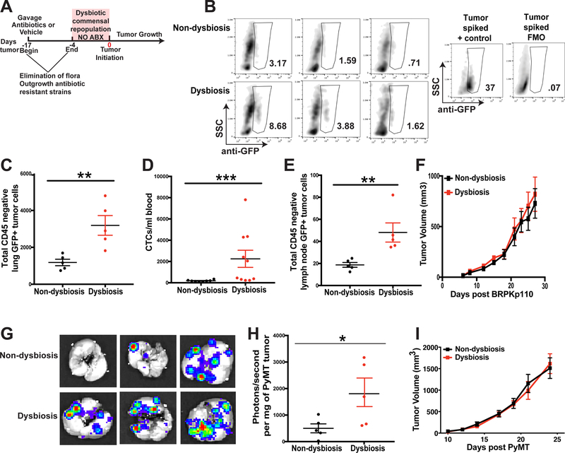
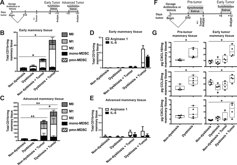
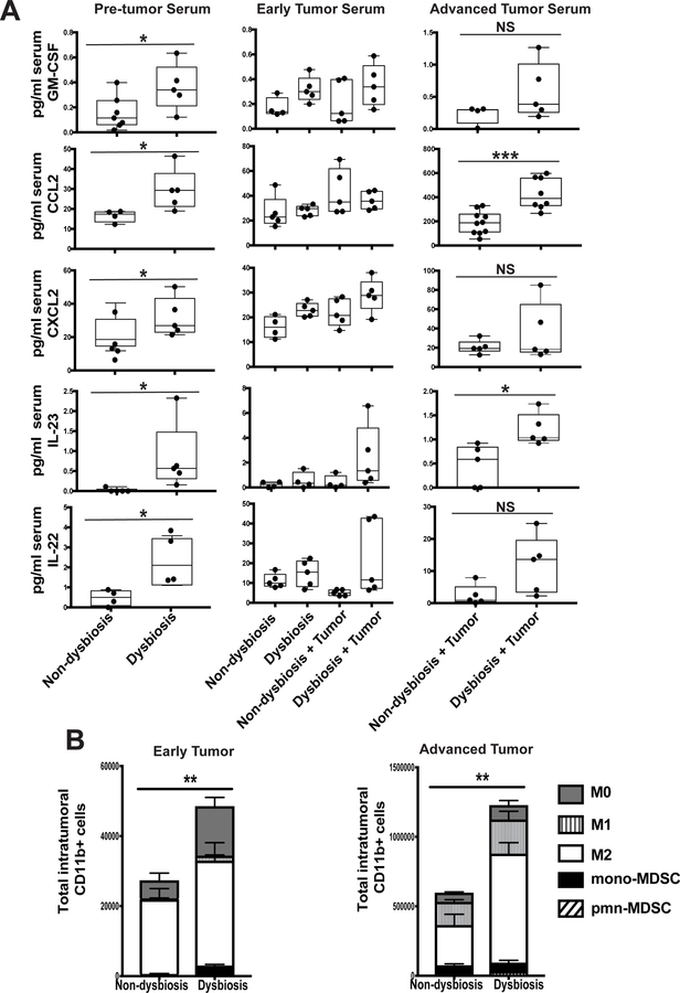
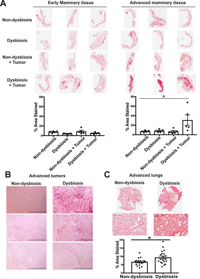
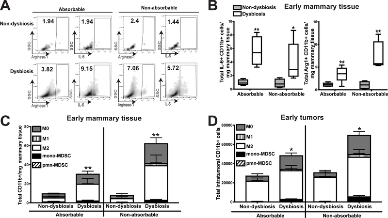
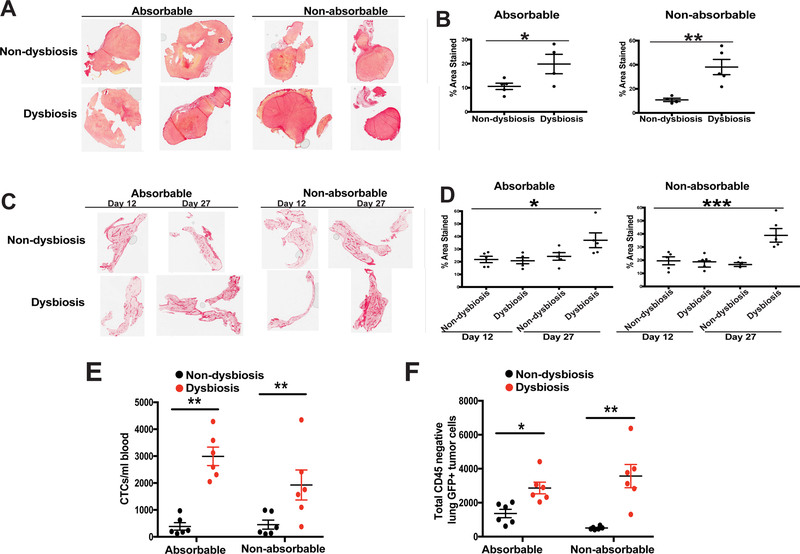
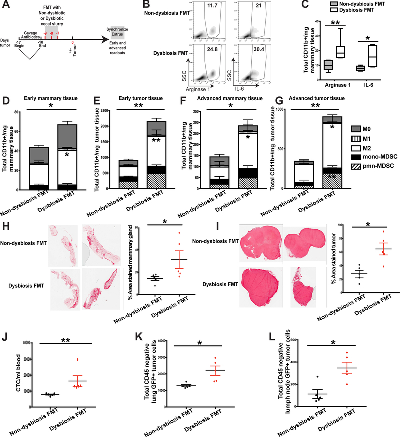
It is unknown why some patients with hormone receptor-positive (HR+) breast cancer present with more aggressive and invasive disease. Metastatic dissemination occurs early in disease and is facilitated by cross-talk between the tumor and tissue environment, suggesting that undefined host-intrinsic factors enhance early dissemination and the probability of developing metastatic disease. Here, we have identified commensal dysbiosis as a host-intrinsic factor associated with metastatic dissemination. Using a mouse model of HR+ mammary cancer, we demonstrate that a preestablished disruption of commensal homeostasis results in enhanced circulating tumor cells and subsequent dissemination to the tumor-draining lymph nodes and lungs. Commensal dysbiosis promoted early inflammation within the mammary gland that was sustained during HR+ mammary tumor progression. Furthermore, dysbiosis enhanced fibrosis and collagen deposition both systemically and locally within the tumor microenvironment and induced significant myeloid infiltration into the mammary gland and breast tumor. These effects were recapitulated both by directly targeting gut microbes using nonabsorbable antibiotics and by fecal microbiota transplantation of dysbiotic cecal contents, demonstrating the direct impact of gut dysbiosis on mammary tumor dissemination. This study identifies dysbiosis as a preexisting, host-intrinsic regulator of tissue inflammation, myeloid recruitment, fibrosis, and dissemination of tumor cells in HR+ breast cancer. SIGNIFICANCE: Identification of commensal dysbiosis as a host-intrinsic factor mediating evolution of metastatic breast cancer allows for development of interventions or diagnostic tools for patients at highest risk for developing metastatic disease.See related commentary by Ingman, p. 3539.


Conflict of interest statement

Conflict of interest disclosure statement: The authors declare no potential conflicts of interest.

Figures

Figure 1
Figure 1. Commensal dysbiosis enhances mammary tumor cell dissemination.
A. Experimental design for antibiotic-induced dysbiosis and tumor initiation. C57BL/6 mice were orally gavaged for 14 days with a broad-spectrum cocktail of antibiotics or an equal volume of water as a vehicle control. Gavage was ceased four days prior to tumor initiation in the fourth mammary fat pad. Tumor size was measured by calipers every 2–3 days after reaching a palpable size. B. Representative plots demonstrating GFP+ tumor cell quantitation in the lungs. The anti-GFP gate was chosen based upon fluorescence minus one (FMO) controls and a stained lung sample spiked with BRPKp110 tumor cells. Numbers represent percent cells within the anti-GFP gate of total live cells. C-F. C57BL/6 mice were treated as described in Fig. 1A. 27 days after BRPKp110 tumor initiation, GFP+ tumor cell dissemination was quantified in lung tissue (C), peripheral blood (D), and tumor-draining axillary lymph nodes (E) by flow cytometry. Data is represented as absolute number of GFP+CD45 cells of live, singlet cells. F. Growth kinetics of BRPKp110 mammary tumors. Representative of at least three independent experiments with 5 mice/group. G-I. C57BL/6 mice were treated as described in Fig. 1A. 25 days after PyMT-luciferase tumor initiation, tumor cell dissemination was quantified in lung tissue by bioluminescence. G. Representative images of bioluminescence in lungs from advanced tumor-bearing mice bearing PyMT-luciferase tumors. H. Quantitation of luminescence represented as photons/second normalized to final tumor burden for mice with PyMT tumors. I. Growth kinetics of PyMT mammary tumors. Representative of two independent experiments with 5 mice/group.
Figure 2
Figure 2. Dysbiosis enhances mammary gland inflammation and myeloid cell infiltration.
A. Experimental design for B-E. C57BL/6 mice were orally gavaged for 14 days with a broad-spectrum cocktail of antibiotics or an equal volume of water as a vehicle control. Gavage was ceased four days prior to BRPKp110 tumor initiation into the abdominal mammary fat pad. Mice were euthanized at early (day 12) and advanced (day 27) tumor timepoints. Four days prior to each timepoint, a modified Whitten effect was used to synchronize estrus in these animals. Normal tumor-adjacent mammary glands were harvested, and infiltrating myeloid cell populations were quantitated by flow cytometry at early (B) and advanced (C) timepoints after tumor initiation. All populations were gated on live, singlet, CD45+CD11b+ cells. Numbers represent absolute numbers of cells quantitated using counting beads. M0 macrophages = F4/80+CD86CD206. M1 macrophages = F4/80+CD86+CD206. M2 macrophages = F4/80+CD86CD206+. Monocytic MDSC = Ly6ChiLy6G. Polymorphonuclear MDSC = Ly6CmidLy6G+. Arginase-1 and IL-6 expression were quantitated by intracellular staining in bulk CD45+CD11b+ cells from mammary glands at early (D) and advanced (E) timepoints after tumor initiation. Numbers represent absolute numbers of cells quantitated using counting beads. F. Experimental design for G. Similar to A, C57BL/6 mice were orally gavaged for 14 days with a broad-spectrum cocktail of antibiotics or an equal volume of water as a vehicle control. Gavage was ceased four days prior to BRPKp110 tumor initiation into the abdominal mammary fat pad. Mice were euthanized prior to tumor initiation (pre-tumor; day 0) and at an early point (early tumor; day 12) during tumor progression. Four days prior to each timepoint, a modified Whitten effect was used to synchronize estrus in these animals. Normal tumor-adjacent mammary glands were harvested, and protein levels of CXCL10, CCL2, and CXCL2 were quantitated by multiplex cytokine analysis (G).
Figure 3
Figure 3. Dysbiosis induces early and sustained systemic inflammation and enhances myeloid infiltration into mammary tumors.
C57BL/6 mice were treated as described in Figure 2A. A. Serum was collected from BRPKp110 tumor- and non-tumor bearing mice, with or without established dysbiosis, at various timepoints: prior to tumor initiation (pre tumor serum; day 0), and at early (early tumor serum; day 12) and advanced (advanced tumor serum; day 27) timepoints after tumor initiation. Serum GM-CSF, CCL2, CXCL2, IL-23, and IL-22 were quantitated by multiplex cytokine analysis. B. BRPKp110 mammary tumors were harvested, and infiltrating myeloid cell populations were quantitated by flow cytometry at early and advanced timepoints after tumor initiation. All populations were gated on live, singlet, CD45+CD11b+ cells. Numbers represent absolute numbers of cells quantitated using counting beads. M0 macrophages = F4/80+CD86CD206. M1 macrophages = F4/80+CD86+CD206. M2 macrophages = F4/80+CD86CD206+. Monocytic MDSC = Ly6ChiLy6G. Polymorphonuclear MDSC = Ly6CmidLy6G+.
Figure 4
Figure 4. Dysbiosis enhances both local and systemic fibrosis within normal tumor-adjacent mammary tissue in advanced tumor-bearing animals.
C57BL/6 mice were treated as described in Figure 2A. A. Normal-adjacent mammary glands were harvested from BRPKp110 tumor- and non-tumor bearing mice, with or without established dysbiosis, at early (early mammary tissue; day 12) and advanced (advanced mammary tissue; day 27) timepoints after tumor initiation. BRPKp110 mammary tumors (B) and lungs (C) were harvested from advanced tumor-bearing animals with or without established dysbiosis. All tissues were formalin-fixed and paraffin-embedded, and sections were stained with PicroSirius Red. Quantification of staining intensity was calculated using Image J software.
Figure 5
Figure 5. Direct targeting of gut commensals using non-absorbable antibiotics results in enhanced inflammation and myeloid cell infiltration in dysbiotic mice.
C57BL/6 mice were treated as described in Figure 2A. Half of the antibiotic-treated animals were gavaged with the previously-described antibiotic cocktail, while the other half were gavaged with a cocktail of non-absorbable antibiotics that have minimal absorption from the gut. Non-dysbiotic animals from each group were gavaged with similar volumes of water per the administered antibiotic cocktail. Normal tumor-adjacent mammary glands (A-C) and BRPKp110 tumors (D) were harvested at an early timepoint (12 days) after tumor initiation, and myeloid cell populations were quantitated by flow cytometry. A. Representative plots gating on arginase-1+ and IL-6+ myeloid cells. Numbers represent the frequency of CD11b+ cells positive for each factor. B. Arginase-1 and IL-6 expression quantitated by intracellular staining in bulk CD45+CD11b+ cells from mammary glands. Numbers represent absolute numbers of cells quantitated using counting beads. Characterization of myeloid populations from mammary glands (C) or tumors (D). All populations were gated on live, singlet, CD45+CD11b+ cells. Numbers represent absolute numbers of cells quantitated using counting beads. M0 macrophages = F4/80+CD86CD206. M1 macrophages = F4/80+CD86+CD206. M2 macrophages = F4/80+CD86CD206+. Monocytic MDSC = Ly6ChiLy6G. Polymorphonuclear MDSC = Ly6CmidLy6G+. Representative of two independent experiments with 5 mice/group
Figure 6
Figure 6. Direct targeting of gut commensals using non-absorbable antibiotics results in enhanced fibrosis and tumor cell dissemination in advanced tumor-bearing dysbiotic mice.
C57BL/6 mice were treated as described in Figure 2A. Half of the antibiotic-treated animals were gavaged with the previously-described antibiotic cocktail while the other half were gavaged with a cocktail of non-absorbable antibiotics that have minimal absorption from the gut. Non-dysbiotic animals from each group were gavaged with similar volumes of water per the administered antibiotic cocktail. A. Advanced BRPKp110 mammary tumors and C. normal tumor-adjacent mammary glands at early (day 12) and advanced (day 27) timepoints after tumor initiation were harvested from tumor-bearing mice, with or without established dysbiosis. Tissues were formalin-fixed and paraffin-embedded, and sections were stained with PicroSirius Red. Quantification of staining intensity was calculated using Image J software for both tumors (B) and mammary glands (D). E-F. GFP+ tumor cell dissemination was quantified in peripheral blood (E) and lung tissue (F) by flow cytometry. Data is represented as absolute number of GFP+CD45 cells of live, singlet cells. The anti-GFP gate was chosen based upon FMOs and a stained lung sample spiked with GFP+ BRPKp110 tumor cells. Representative of two independent experiments with 5 mice/group
Figure 7
Figure 7. Fecal microbiota transplantation of dysbiotic cecal contents is sufficient to enhance inflammation, myeloid cell infiltration, fibrosis, and tumor cell dissemination.
A. Experimental design for B-L. C57BL/6 mice were orally gavaged for 7 days with a broad-spectrum cocktail of antibiotics to create a niche for fecal transplantation. Immediately following antibiotic cessation, mice received three consecutive days of oral gavage of cecal slurries collected from dysbiotic or non-dysbiotic mice from day 0, as depicted in Fig. 2A. Mice were then rested for seven days to allow for bacterial engraftment and growth prior to BRPKp110 tumor initiation into the abdominal mammary fat pad. Mice were euthanized at early (day 12) and advanced (day 27) tumor timepoints. Four days prior to each timepoint, a modified Whitten effect was used to synchronize estrus in these animals. B-C. Arginase-1 and IL-6 expression were quantitated by intracellular staining in bulk CD45+CD11b+ cells from mammary glands. B. Representative intracellular staining of arginase-1 and IL-6 from myeloid cells within normal-adjacent mammary glands. Numbers represent percent of total CD11b+ myeloid cells. C. Quantitation of CD11b+ cells producing arginase-1 or IL-6. Numbers represent absolute numbers quantitated using counting beads. D-G. Mammary glands and tumors were harvested at early or advanced tumor timepoints and myeloid cell populations were quantitated by flow cytometry. All populations were gated on live, singlet, CD45+CD11b+ cells. Numbers represent absolute numbers of cells quantitated using counting beads. D. Absolute number of myeloid populations in early mammary glands. E. Absolute number of myeloid populations in early tumors. F. Absolute number of myeloid populations in advanced mammary glands. G. Absolute number of myeloid populations in advanced tumors. M0 macrophages = F4/80+CD86CD206. M1 macrophages = F4/80+CD86+CD206. M2 macrophages = F4/80+CD86CD206+. Monocytic MDSC = Ly6ChiLy6G. Polymorphonuclear MDSC = Ly6CmidLy6G+. Normal-adjacent mammary glands (H) and tumors (I) were harvested from advanced tumor-bearing animals that received either dysbiotic or non-dysbiotic FMT. Tissues were formalin-fixed and paraffin-embedded, and sections were stained with PicroSirius Red. Quantification of staining intensity was calculated using Image J software. Representative of two independent experiments with 5 mice/group. J-L. GFP+ tumor cell dissemination was quantified in peripheral blood (J) lung tissue (K) and distal axillary lymph nodes (L) by flow cytometry. Data is represented as absolute number of GFP+CD45 cells of live, singlet cells. The anti-GFP gate was chosen based upon FMOs and a stained lung sample spiked with GFP+ BRPKp110 tumor cells. Representative of two independent experiments with 5 mice/group.

Comment in

References

    1. Burstein HJ, Cirrincione CT, Barry WT, Chew HK, Tolaney SM, Lake DE, et al. Endocrine therapy with or without inhibition of epidermal growth factor receptor and human epidermal growth factor receptor 2: a randomized, double-blind, placebo-controlled phase III trial of fulvestrant with or without lapatinib for postmenopausal women with hormone receptor-positive advanced breast cancer-CALGB 40302 (Alliance). J Clin Oncol 2014;32:3959–66. 10.1200/JCO.2014.56.7941. Epub 2014 Oct 27. - DOI - PMC - PubMed
    1. DeSantis CE, Ma J, Goding Sauer A, Newman LA, Jemal A. Breast cancer statistics, 2017, racial disparity in mortality by state. CA Cancer J Clin 2017;67:439–48. 10.3322/caac.21412. Epub 2017 Oct 3. - DOI - PubMed
    1. Song F, Zhang J, Li S, Wu J, Jin T, Qin J, et al. ER-positive breast cancer patients with more than three positive nodes or grade 3 tumors are at high risk of late recurrence after 5-year adjuvant endocrine therapy. Onco Targets Ther 2017;10:4859–4867.: 10.2147/OTT.S142698. eCollection 2017. - DOI - PMC - PubMed
    1. Colzani E, Johansson AL, Liljegren A, Foukakis T, Clements M, Adolfsson J, et al. Time-dependent risk of developing distant metastasis in breast cancer patients according to treatment, age and tumour characteristics. Br J Cancer 2014;110:1378–84 - PMC - PubMed
    1. Hosseini H, Obradovic MM, Hoffmann M, Harper KL, Sosa MS, Werner-Klein M, et al. Early dissemination seeds metastasis in breast cancer. Nature 2016 - PMC - PubMed