. 2019 May 7;11(9):2762-2786.
doi: 10.18632/aging.101948.

MiR-191 inhibit angiogenesis after acute ischemic stroke targeting VEZF1

Affiliations

MiR-191 inhibit angiogenesis after acute ischemic stroke targeting VEZF1

Kang Du et al. Aging (Albany NY). .

Abstract

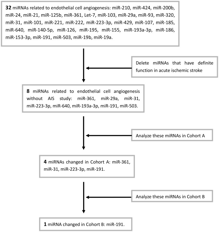
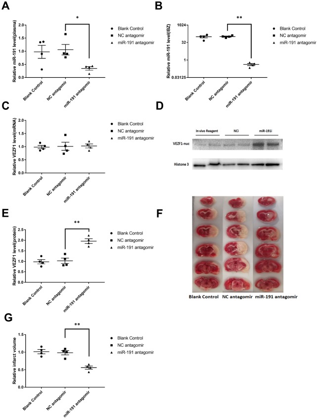
Acute ischemic stroke (AIS) is a major public health problem in China. Impaired angiogenesis plays crucial roles in the development of ischemic cerebral injury. Recent studies have identified that microRNAs (miRNAs) are important regulators of angiogenesis, but little is known the exact effects of angiogenesis-associated miRNAs in AIS. In the present study, we detected the expression levels of angiogenesis-associated miRNAs in AIS patients, middle cerebral artery occlusion (MCAO) rats, and oxygen-glucose deprivation/reoxygenation (OGD/R) human umbilical vein endothelial cells (HUVECs). MiR-191 was increased in the plasma of AIS patients, OGD/R HUVECs, and the plasma and brain of MCAO rats. Over-expression of miR-191 promoted apoptosis, but reduced the proliferation, migration, tube-forming and spheroid sprouting activity in HUVECs OGD/R model. Mechanically, vascular endothelial zinc finger 1 (VEZF1) was identified as the direct target of miR-191, and could be regulated by miR-191 at post-translational level. In vivo studies applying miR-191 antagomir demonstrated that inhibition of miR-191 reduced infarction volume in MCAO rats. In conclusion, our data reveal a novel role of miR-191 in promoting ischemic brain injury through inhibiting angiogenesis via targeting VEZF1. Therefore, miR-191 may serve as a biomarker or a therapeutic target for AIS.

Keywords: VEZF1; acute stroke; angiogenesis factor; miR-191.


Conflict of interest statement

CONFLICTS OF INTEREST: The authors have announced no conflicts of interest.

Figures

Figure 1
Figure 1
Relative miRNAs levels. Expression levels of miRNAs in Cohort A+B (n=18) (A) miR-31, (B) miR-191, (C) miR-223-3p, (D) miR-361; (E) Expression level of miR-191 in rat MCAO plasma after 24h reperfusion (n=12); (F) Expression level of miR-191 in rat MCAO plasma after 48h reperfusion (n=12); (G) Expression level of miR-191 in rat MCAO brains (n=12); (H) Expression level of miR-191 in OGD HUVECs (n=6). Means ± SEM. * P< 0.05,** P< 0.01 vs. NCm or NCi.
Chart 1
Chart 1. Screening process of miRNAs.
Figure 2
Figure 2
MiR-191 transfection efficiency and cell proliferation. (A, B) Expression level of miR-191 (in fold of NCm or percentage of NCi) in HUVECs that were transfected with miR-191 mimic (A) or miR-191 inhibitor (B) as assessed by real-time PCR (n=6 per group); (C, D) Proliferation (percentage of NCm or NCi) of HUVECs transfected with miR-191 mimic (C) or miR-191 inhibitor (D) as assessed by CCK-8 assay (n =6 per group). After transfection, the cells were reseeded into 96-well plates and incubated for another 48 h. Means ± SEM. ** P< 0.01 vs. NCm or NCi.
Figure 3
Figure 3
Cell apoptosis and cell cycle. (A, B) Cell apoptosis analysis of HUVECs transfected with miR-191 mimic (A) or miR-191 inhibitor (B)as assessed by flow cytometry; (C, D) Percentage of apoptosis HUVECs transfected with miR-191 mimic (C) or miR-191 inhibitor (D) (n =6 per group); (E, F) Cell cycle analysis of HUVECs transfected with miR-191 mimic (E) or miR-191 inhibitor (F) as assessed by flow cytometry; (G, H) Percentage of HUVECs transfected with miR-191 mimic (G) or miR-191 inhibitor (H) in different stages (n=6 per group). Means ± SEM.,** P< 0.01 vs. NCm or NCi.
Figure 4
Figure 4
MiR-191 inhibited cell migration. (A, B) Phase contrast microscopic images of HUVECs at 0, 6, and 12 h after scratching. The cells were transfected with miR-191 mimic (A), miR-191 inhibitor (B), or the corresponding scrambled NCm (A) and NCi (B). Black lines indicate the wound area. Scale bars, 100 μm. (C, D) Size of wound area (percentage of 0 h) created by scratching HUVECs transfected with miR-191 mimic (C) or miR-191 inhibitor (D) (n =6 per group). (E, F) Phase contrast microscopic images of HUVECs migrated and attached to the bottom membrane of a transwell. The cells were transfected with miR-191 mimic (E), miR-191 inhibitor (F), or the corresponding scrambled NCm (E) and NCi (F). Scale bars, 20μm. (G, H) Number of migrated HUVECs (percentage of NCm or NCi) transfected with miR-191 mimic (G) or miR-191 inhibitor (H) (n =6 per group). Means ± SEM. * P< 0.05,** P< 0.01 vs. NCm or NCi.
Figure 5
Figure 5
miR-191 inhibited tube formation and spheroid sprouting. (A, B) Phase-contrast microscopic images of tubeforming HUVECs that were transfected with miR-191 mimic (A), miR-191 inhibitor (B), or the corresponding scrambled NCm (A) and NCi (B). Scale bars, 20μm. (C, D) Tube formation of HUVECs (percentage of NCm or NCi) transfected with miR-191 mimic (C) or miR-191 inhibitor (D) as assessed by tube formation assay (n =6 per group). (E, F) Phase-contrast microscopic images of sprouting HUVECs spheroids. HUVECs were transfected with miR-191 mimic (E), miR-191 inhibitor (F), or the corresponding scrambled NCm (E) and NCi (F). Scale bars, 20μm. (G, H) Sprouting coverage area of HUVECs (percentage of NCm or NCi) transfected with miR-191 mimic (G) or miR-191 inhibitor (H) as assessed by spheroid sprouting assay (n = 6 per group). Means ± SEM. ** P< 0.01 vs. NCm or NCi.
Figure 6
Figure 6
Regulation of VEZF1 by miR-191. (A) MiR-191 and its putative binding sequence in the 3'-UTR of VEZF1. (B) Real-time PCR analysis of VEZF1 expression in HUVECs by miR-191 interference. (n =6 per group); (C, D) Western blot analysis of VEZF1 expression in HUVECs transfected by miR-191. (E) Construction of Plasmid; (F) Luciferase activities were measured to evaluate the binding of miR-191 to the candidate binding sequence of VEZF1 after transfection of miR-191 mimic or NCm. Means ± SEM. * P< 0.05,** P< 0.01 vs. NCm or NCi.
Figure 7
Figure 7
mRNA levels of Targets of VEZF1. Real-time PCR for EDN1 (A, B), MMP1 (C, D), STMN1 (E, F), CITED2 (G, H), MMP2 (I, J), and MMP9 (K, L) mRNA of HUVECs transfected with miR-191 mimic or miR-191 inhibitor (n = 6 per group). Means ± SEM. * P< 0.05,** P< 0.01 vs. NCm or NCi.
Figure 8
Figure 8
Inhibition of miR-191 reduced infarction volume of MCAO rats after the injection of miR-191 antagomir compared to NC antagomir and blank control. Relative miR-191 levels in (A) plasma and (B) IBZ (n =4 per group); (C) Real-time PCR analysis of VEZF1 expression in IBZ (n =4 per group); (D, E) Western blot analysis of VEZF1 expression in IBZ (n =4 per group); (F, G) Relative infarct volume of MCAO rats (n =4 per group). Means ± SEM. * P< 0.05,** P< 0.01 vs. NC antagomir.
Figure 9
Figure 9
miR-191 reduces the nucleoprotein VEZF1, resulting in up-regulation of CITED2 and down-regulation of MMP-1, STMN1 mRNA. This, in turn, suppresses HUVEC proliferation and migration and thus prevents endothelial tube formation and spheroid sprouting. Moreover, miR-191 suppresses the expression of EDN1 which functions as maintaining vascular tension.

References

    1. Wei JW, Heeley EL, Wang JG, Huang Y, Wong LK, Li Z, Heritier S, Arima H, Anderson CS, and ChinaQUEST Investigators. Comparison of recovery patterns and prognostic indicators for ischemic and hemorrhagic stroke in China: the ChinaQUEST (QUality Evaluation of Stroke Care and Treatment) Registry study. Stroke. 2010; 41:1877–83. 10.1161/STROKEAHA.110.586909 - DOI - PubMed
    1. Powers WJ, Rabinstein AA, Ackerson T, Adeoye OM, Bambakidis NC, Becker K, Biller J, Brown M, Demaerschalk BM, Hoh B, Jauch EC, Kidwell CS, Leslie-Mazwi TM, et al., and American Heart Association Stroke Council. 2018 Guidelines for the Early Management of Patients With Acute Ischemic Stroke: A Guideline for Healthcare Professionals From the American Heart Association/American Stroke Association. Stroke. 2018; 49:e46–110. 10.1161/STR.0000000000000158 - DOI - PubMed
    1. Licata G, Tuttolomondo A, Corrao S, Di Raimondo D, Fernandez P, Caruso C, Avellone G, Pinto A. Immunoinflammatory activation during the acute phase of lacunar and non-lacunar ischemic stroke: association with time of onset and diabetic state. Int J Immunopathol Pharmacol. 2006; 19:639–46. 10.1177/039463200601900320 - DOI - PubMed
    1. Tuttolomondo A, Di Sciacca R, Di Raimondo D, Pedone C, La Placa S, Pinto A, Licata G. Effects of clinical and laboratory variables and of pretreatment with cardiovascular drugs in acute ischaemic stroke: a retrospective chart review from the GIFA study. Int J Cardiol. 2011; 151:318–22. 10.1016/j.ijcard.2010.06.005 - DOI - PubMed
    1. Di Raimondo D, Tuttolomondo A, Buttà C, Miceli S, Licata G, Pinto A. Effects of ACE-inhibitors and angiotensin receptor blockers on inflammation. Curr Pharm Des. 2012; 18:4385–413. 10.2174/138161212802481282 - DOI - PubMed

Publication types

MeSH terms