. 2019 May 7;9(1):7008.
doi: 10.1038/s41598-019-43453-7.

Increased elasticity of melanoma cells after low-LET proton beam due to actin cytoskeleton rearrangements

Affiliations

Increased elasticity of melanoma cells after low-LET proton beam due to actin cytoskeleton rearrangements

Katarzyna Jasińska-Konior et al. Sci Rep. .

Abstract

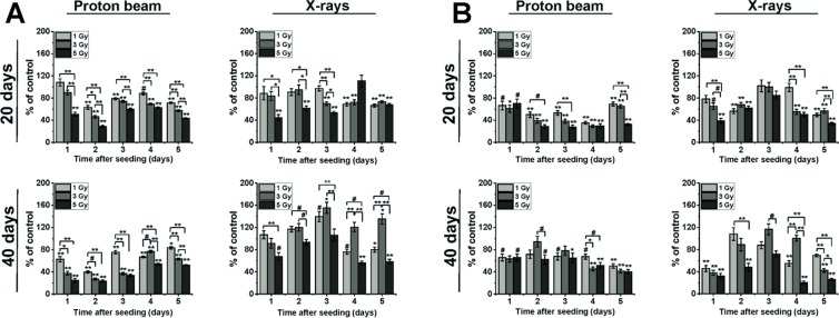
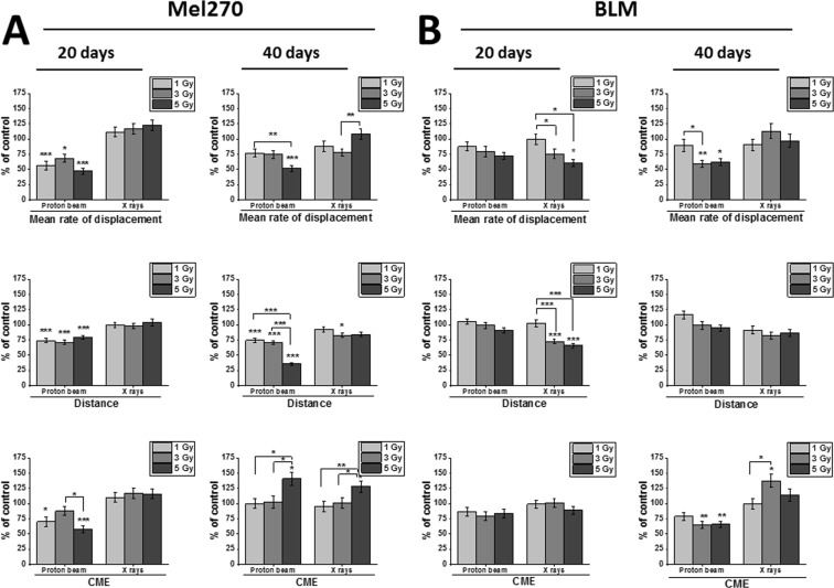
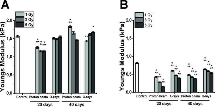
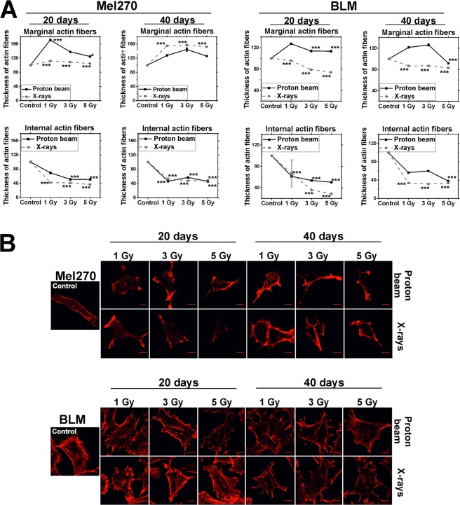
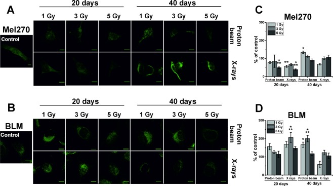
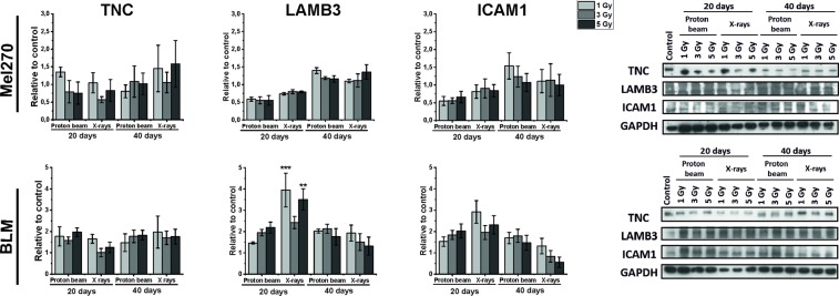
Cellular response to non-lethal radiation stress include perturbations in DNA repair, angiogenesis, migration, and adhesion, among others. Low-LET proton beam radiation has been shown to induce somewhat different biological response than photon radiation. For example, we have shown that non-lethal doses of proton beam radiation inhibited migration of cells and that this effect persisted long-term. Here, we have examined cellular elasticity and actin cytoskeleton organization in BLM cutaneous melanoma and Mel270 uveal melanoma cells. Proton beam radiation increased cellular elasticity to a greater extent than X-rays and both types of radiation induced changes in actin cytoskeleton organization. Vimentin level increased in BLM cells after both types of radiation. Our data show that cell elasticity increased substantially after low-LET proton beam and persisted long after radiation. This may have significant consequences for the migratory properties of melanoma cells, as well as for the cell susceptibility to therapy.


Conflict of interest statement

The authors declare no competing interests.

Figures

Figure 1
Figure 1
Cell counting of Mel270 (A) and BLM (B) cells performed 20 and 40 days after treatment with proton beam radiation or X-rays. Cells were irradiated, cultured at standard conditions and then seeded at 20 and 40 days post-treatment and the assay was conducted during five next days. Experiment was performed in triplicate and for every repetition, cells from 4 wells were counted. Results are presented as the percent of the untreated control. Mean values ± SEM. #p < 0.05; *p < 0.01; **p < 0.001.
Figure 2
Figure 2
Cellular migration properties of Mel270 (A) and BLM (B) cells treated with proton beam radiation or X rays. Individual cell movements were evaluated at 20 and 40 days after irradiation with either proton beam or X-rays and three parameters were calculated: ‘Mean rate of displacement’, i.e. the distance from the starting point direct to the cell's final position/time of recording; ‘Distance’, i.e. the total cell trajectory (μm) and CME (coefficient of movement efficiency), i.e. the ratio of cell displacement to the cell trajectory length. Mean values presented as percent of control; *p < 0.05, **p < 0.01, ***p < 0.001. Data partially presented in Jasinska et al..
Figure 3
Figure 3
Impact of proton beam radiation and X-rays on the elastic properties of Mel270 (A) and BLM cells (B). Mean of Young modulus measured from 325 cells ± SEM values; *p < 0.05; **p < 0.01; ***p < 0.001, against untreated control.
Figure 4
Figure 4
(A) AFM amplitude images of the BLM and Mel270 cells. (B) Confocal microscopy images of the same cells stained for actin (red), vimentin (green) and nucleus (blue). Scale bars in all images represent 10 um. Cells were treated with 3 Gy of either proton beam or X-rays and cultured for 20 days.
Figure 5
Figure 5
(A) Thickness of actin fibers in Mel270 and BLM cells treated with low-LET proton beam radiation or X-rays. Measurements of 15 cells in each experimental group were taken at 20 and 40 days following radiation. Two types of fibers were measured: marginal and internal also called stress fibers with 15 measurements for each type of fibers per cell. Results are presented as the percent of the untreated control. Mean values ± SEM. *p < 0.05; **p < 0.01; ***p < 0.001. (B) Representative images of actin cytoskeleton stained in Mel270 and BLM cells after treatment with low-LET proton beam radiation or X-rays. Cells were stained with phalloidin conjugated with Alexa 546. Scale bar: 10 μm.
Figure 6
Figure 6
Representative images of vimentin stained in Mel270 (A) and BLM (B) cells after treatment with proton beam radiation or X-rays. The corrected total cell fluorescence (CTCF) was measured in Mel270 (C) and BLM (D) cells. For each experimental group 15 cells were measured. Results are presented as the percent of the untreated control. Mean values ± SEM. *p < 0.05; **p < 0.01; ***p < 0.001. Scale bar: 10 µm.
Figure 7
Figure 7
TNC, LAMB3 and ICAM1 protein expression determined with Western Blot in Mel270 and BLM cells after treatment with different doses (1, 3, 5 Gy) of proton beam or X rays. Cells were lysed 20 and 40 days after irradiation. Protein bands were densitrometrically analyzed and adjusted against GAPDH. Results represent average of 3 independent experiments. Mean ± SEM. *p < 0.05; **p < 0.01; ***p < 0.001. Samples were analyzed together as indicated by the frames (only TNC control was taken from another blot, see Suppl. Fig. A3).

Similar articles

Cited by

References

    1. Verschueren KMS, et al. Long-term outcomes of eye-conserving treatment with Ruthenium106 brachytherapy for choroidal melanoma. Radiother. Oncol. 2010;95:332–338. doi: 10.1016/j.radonc.2010.03.023. - DOI - PubMed
    1. Allen AM, et al. An evidence based review of proton beam therapy: the report of ASTRO’s emerging technology committee. Radiother. Oncol. 2012;103:8–11. doi: 10.1016/j.radonc.2012.02.001. - DOI - PubMed
    1. Gragoudas ES. The Bragg Peak of proton Beams for Treatment of Uveal melanoma. Int. Ophthalmol. Clin. 1980;20:123–133. - PubMed
    1. Egger E, et al. Eye retention after proton beam radiotherapy for uveal melanoma. Int. J. Radiat. Oncol. 2003;55:867–880. doi: 10.1016/S0360-3016(02)04200-1. - DOI - PubMed
    1. Collaborative Ocular Melanoma Study Group The COMS randomized trial of iodine 125 brachytherapy for choroidal melanoma: V. Twelve-year mortality rates and prognostic factors: COMS report No. 28. Arch Ophthalmol. 2006;12:1684–93. - PubMed

Publication types

MeSH terms