. 2019 May 7;9(1):7030.
doi: 10.1038/s41598-019-43417-x.

Increased LIGHT expression and activation of non-canonical NF-κB are observed in gastric lesions of MyD88-deficient mice upon Helicobacter felis infection

Affiliations

Increased LIGHT expression and activation of non-canonical NF-κB are observed in gastric lesions of MyD88-deficient mice upon Helicobacter felis infection

Raquel Mejías-Luque et al. Sci Rep. .

Abstract

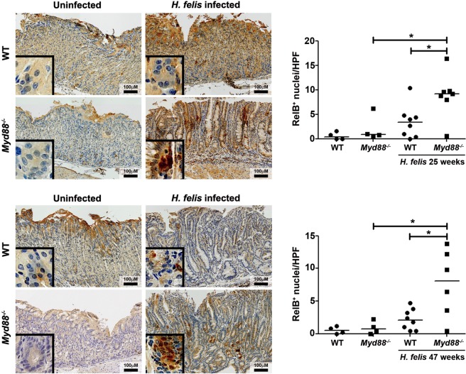
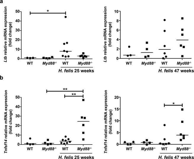
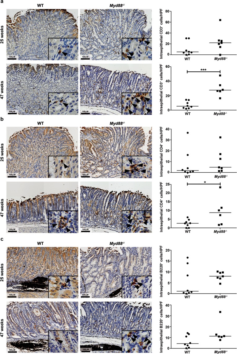
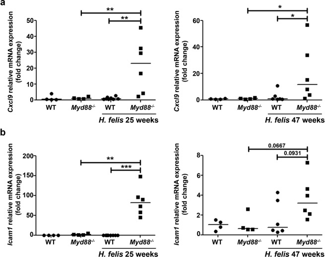
Helicobacter pylori infection induces a number of pro-inflammatory signaling pathways contributing to gastric inflammation and carcinogenesis. Among those, NF-κB signaling plays a pivotal role during infection and malignant transformation of the gastric epithelium. However, deficiency of the adaptor molecule myeloid differentiation primary response 88 (MyD88), which signals through NF-κB, led to an accelerated development of gastric pathology upon H. felis infection, but the mechanisms leading to this phenotype remained elusive. Non-canonical NF-κB signaling was shown to aggravate H. pylori-induced gastric inflammation via activation of the lymphotoxin β receptor (LTβR). In the present study, we explored whether the exacerbated pathology observed in MyD88-deficient (Myd88-/-) mice was associated with aberrant activation of non-canonical NF-κB. Our results indicate that, in the absence of MyD88, H. felis infection enhances the activation of non-canonical NF-κB that is associated with increase in Cxcl9 and Icam1 gene expression and CD3+ lymphocyte recruitment. In addition, activation of signal transducer and activator of transcription 3 (STAT3) signaling was higher in Myd88-/- compared to wild type (WT) mice, indicating a link between MyD88 deficiency and STAT3 activation in response to H. felis infection. Thereby, MyD88 deficiency results in accelerated and aggravated gastric pathology induced by Helicobacter through activation of non-canonical NF-κB.


Conflict of interest statement

The authors declare no competing interests.

Figures

Figure 1
Figure 1
Non-canonical NF-κB signaling is enhanced in Myd88−/− mice upon H. felis infection. Representative images of RelB expression in gastric tissue samples of wild type (WT) and Myd88−/− mice control or infected with H. felis for 25 or 47 weeks. Quantification of RelB+ cells per high power field (HPF) (20x magnification) is shown. *p ≤ 0.05, **p ≤ 0.01. T-test. Each dot represents one mouse. Horizontal bars indicate medians.
Figure 2
Figure 2
LIGHT is the main ligand inducing non-canonical NF-κB upon H. felis infection. Relative mRNA expression levels of LTb (a) and Tnfsf14 (b) in the stomach of control and H. felis-infected mice at 25 and 47 weeks post-infection. Ct values were normalized to Gadph. *p ≤ 0.05, **p ≤ 0.01. Kruskal-Wallis test. Each dot represents one mouse. Horizontal bars indicate medians.
Figure 3
Figure 3
Recruitment of T and B lymphocytes into the stomach of H. felis-infected mice. Murine stomach samples of WT and Myd88−/− mice infected with H. felis were stained for CD3 (a), CD4 (b), and B220 (c) by immunohistochemistry. Representative images are shown as well as quantification of CD3, CD4, and B220 positive cells (brown) per high power field (20x magnification). ***p ≤ 0.001. Mann-Whitney U test. Each dot represents one mouse. Horizontal bars indicate medians.
Figure 4
Figure 4
Cxcl9 and Icam1 are up-regulated in Myd88−/− mice upon H. felis infection. Cxcl9 (a) and Icam1 (b) mRNA levels in control and H. felis-infected mice was determined by real-time PCR. Ct values were normalized to Gadph. *p ≤ 0.05, **p ≤ 0.01, ***p ≤ 0.001. Kruskal-Wallis test. Each dot represents one mouse. Horizontal bars indicate medians.
Figure 5
Figure 5
H. felis infection enhances STAT3 activation in Myd88−/− mice. Representative images of p-STAT3 expression in the stomach of WT and Myd88−/− mice infected with H. felis for 25 and 47 weeks. Quantification of p-STAT3+ cells per high power field (HPF) (20x magnification) is shown. *p ≤ 0.05. Mann-Whitney U test. Each dot represents one mouse. Horizontal bars indicate medians.

References

    1. Moyat M, Velin D. Immune responses to Helicobacter pylori infection. World journal of gastroenterology: WJG. 2014;20:5583–5593. doi: 10.3748/wjg.v20.i19.5583. - DOI - PMC - PubMed
    1. Ernst M, Thiem S, Nguyen PM, Eissmann M, Putoczki TL. Epithelial gp130/Stat3 functions: an intestinal signaling node in health and disease. Semin Immunol. 2014;26:29–37. doi: 10.1016/j.smim.2013.12.006. - DOI - PubMed
    1. Menheniott TR, Judd LM, Giraud AS. STAT3: a critical component in the response to Helicobacter pylori infection. Cellular microbiology. 2015;17:1570–1582. doi: 10.1111/cmi.12518. - DOI - PubMed
    1. Jenkins BJ, et al. Hyperactivation of Stat3 in gp130 mutant mice promotes gastric hyperproliferation and desensitizes TGF-beta signaling. Nature medicine. 2005;11:845–852. doi: 10.1038/nm1282. - DOI - PubMed
    1. Backert S, Naumann M. What a disorder: proinflammatory signaling pathways induced by Helicobacter pylori. Trends in microbiology. 2010;18:479–486. doi: 10.1016/j.tim.2010.08.003. - DOI - PubMed

Publication types

MeSH terms