. 2019 Apr 1:2019:6978303.
doi: 10.1155/2019/6978303. eCollection 2019.

Induced Pluripotent Stem Cell Derivation and Ex Vivo Gene Correction Using a Mucopolysaccharidosis Type 1 Disease Mouse Model

Affiliations

Induced Pluripotent Stem Cell Derivation and Ex Vivo Gene Correction Using a Mucopolysaccharidosis Type 1 Disease Mouse Model

Toshio Miki et al. Stem Cells Int. .

Abstract

Mucopolysaccharidosis type 1 (MPS-1), also known as Hurler's disease, is a congenital metabolic disorder caused by a mutation in the alpha-L-iduronidase (IDUA) gene, which results in the loss of lysosomal enzyme function for the degradation of glycosaminoglycans. Here, we demonstrate the proof of concept of ex vivo gene editing therapy using induced pluripotent stem cell (iPSC) and CRISPR/Cas9 technologies with MPS-1 model mouse cell. Disease-affected iPSCs were generated from Idua knockout mouse embryonic fibroblasts, which carry a disrupting neomycin-resistant gene cassette (Neor) in exon VI of the Idua gene. Double guide RNAs were used to remove the Neor sequence, and various lengths of donor templates were used to reconstruct the exon VI sequence. A quantitative PCR-based screening method was used to identify Neor removal. The sequence restoration without any indel mutation was further confirmed by Sanger sequencing. After induced fibroblast differentiation, the gene-corrected iPSC-derived fibroblasts demonstrated Idua function equivalent to the wild-type iPSC-derived fibroblasts. The Idua-deficient cells were competent to be reprogrammed to iPSCs, and pluripotency was maintained through CRISPR/CAS9-mediated gene correction. These results support the concept of ex vivo gene editing therapy using iPSC and CRISPR/Cas9 technologies for MPS-1 patients.


Figures

Figure 1
Figure 1
Schematic representation of the Idua−/− iPSC derivation and gene correction. Idua−/− mouse embryonic fibroblasts were reprogrammed with STEMCCA lentivirus. The disrupting neomycin resistance gene (Neor) was removed, and the Idua gene was corrected with CRISPR/Cas9 gene editing technology.
Figure 2
Figure 2
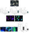
Idua−/− mouse iPSC derivation and validation. (a) Phase contrast image of miPSC colonies. (b) Stem cell marker gene expression in three different miPSC lines. Mouse embryonic stem cell (mES) as control. (c) Immunofluorescent staining for stem cell markers, SSEA-1 and Oct4. Nuclear counter staining with DAPI. (d) Immunofluorescent staining demonstrated lineage-specific marker protein-positive cells. (e) Biochemical analyses demonstrate IDUA activity in wild-type (WT) and IDUA knockout (IDUA KO) fibroblasts and iPSCs derived from each cell line.
Figure 3
Figure 3
CRISPR gene editing and validations. (a) Schematic representation of CRISPR gene editing strategy. (b) Flow cytometric analysis of CRISPR/Cas9-OFP vector transfection efficiency. GFP channel was used to detect autofluorescence of dead cells. (c) Stem cell marker gene expression in two gene-edited miPSC lines. (d) Identification of three germ layers in teratomas. Red arrows indicate typical histological feature of each lineage. (e) Biochemical analyses to validate Idua enzyme function restoration. P < 0.05 and ∗∗ P < 0.005. n.s: no significant difference using one-way ANOVA with Bonferroni post hoc analysis and Dunnett's multiple comparison. (f) The retention of restored Idua enzyme activity was confirmed after inducing cell differentiation (Fib.: fibroblast-like cells, Ne./Sp.: spontaneously induced neuronal-shape cells). P < 0.05 and ∗∗ P < 0.005. n.s: no significant difference using one-way ANOVA with Bonferroni post hoc analysis and Dunnett's multiple comparison.

References

    1. Terlato N. J., Cox G. F. Can mucopolysaccharidosis type I disease severity be predicted based on a patient’s genotype? A comprehensive review of the literature. Genetics in Medicine. 2003;5(4):286–294. doi: 10.1097/01.GIM.0000078027.83236.49. - DOI - PubMed
    1. Venturi N., Rovelli A., Parini R., et al. Molecular analysis of 30 mucopolysaccharidosis type I patients: evaluation of the mutational spectrum in Italian population and identification of 13 novel mutations. Human Mutation. 2002;20(3):231–231. doi: 10.1002/humu.9051. - DOI - PubMed
    1. Qin Y., Gao W. Q. Concise review: patient-derived stem cell research for monogenic disorders. Stem Cells. 2016;34(1):44–54. doi: 10.1002/stem.2112. - DOI - PubMed
    1. Pandey V. K., Tripathi A., Bhushan R., Ali A., Dubey P. K. Application of CRISPR/Cas9 genome editing in genetic disorders: a systematic review up to date. Journal of Genetic Syndromes & Gene Therapy. 2017;8(2) doi: 10.4172/2157-7412.1000321. - DOI
    1. Jinek M., Chylinski K., Fonfara I., Hauer M., Doudna J. A., Charpentier E. A programmable dual-RNA-guided DNA endonuclease in adaptive bacterial immunity. Science. 2012;337(6096):816–821. doi: 10.1126/science.1225829. - DOI - PMC - PubMed

LinkOut - more resources