. 2019 Apr 1:2019:8940365.
doi: 10.1155/2019/8940365. eCollection 2019.

Improved Efficiency of Cardiomyocyte-Like Cell Differentiation from Rat Adipose Tissue-Derived Mesenchymal Stem Cells with a Directed Differentiation Protocol

Affiliations

Improved Efficiency of Cardiomyocyte-Like Cell Differentiation from Rat Adipose Tissue-Derived Mesenchymal Stem Cells with a Directed Differentiation Protocol

Blanca Rebeca Ibarra-Ibarra et al. Stem Cells Int. .

Abstract

Cell-based therapy has become a resource for the treatment of cardiovascular diseases; however, there are some conundrums to achieve. In vitro cardiomyocyte generation could be a solution for scaling options in clinical applications. Variability on cardiac differentiation in previously reported studies from adipose tissue-derived mesenchymal stem cells (ASCs) and the lack of measuring of the cardiomyocyte differentiation efficiency motivate the present study. Here, we improved the ASC-derived cardiomyocyte-like cell differentiation efficiency with a directed cardiomyocyte differentiation protocol: BMP-4 + VEGF (days 0-4) followed by a methylcellulose-based medium with cytokines (IL-6 and IL-3) (days 5-21). Cultures treated with the directed cardiomyocyte differentiation protocol showed cardiac-like cells and "rosette-like structures" from day 7. The percentage of cardiac troponin T- (cTnT-) positive cells was evaluated by flow cytometry to assess the cardiomyocyte differentiation efficiency in a quantitative manner. ASCs treated with the directed cardiomyocyte differentiation protocol obtained a differentiation efficiency of up to 44.03% (39.96%±3.78) at day 15 without any enrichment step. Also, at day 21 we observed by immunofluorescence the positive expression of early, late, and cardiac maturation differentiation markers (Gata-4, cTnT, cardiac myosin heavy chain (MyH), and the sarcoplasmic/endoplasmic reticulum Ca2+ ATPase (SERCa2)) in cultures treated with the directed cardiomyocyte differentiation protocol. Unlike other protocols, the use of critical factors of embryonic cardiomyogenesis coupled with a methylcellulose-based medium containing previously reported cardiogenic cytokines (IL-6 and IL-3) seems to be favorable for in vitro cardiomyocyte generation. This novel efficient culture protocol makes ASC-derived cardiac differentiation more efficient. Further investigation is needed to identify an ASC-derived cardiomyocyte surface marker for cardiac enrichment.


Figures
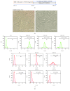
Figure 1
Figure 1
(a) Schematic outline of the directed cardiomyocyte differentiation protocol (days). Only growth factors and the combination of growth factors plus MethoCult M3534 conditions are shown. Undifferentiated ASCs. (b) A spindle fibroblast-like cell morphology was assessed by inverted microscope imaging (scale bar, 100 μm). (c) Flow cytometry histograms for stem cell surface markers versus forward scatter (FSC), M1 (percentage of positive cells (%), mean ± SD, n = 3).
Figure 2
Figure 2
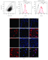
Undifferentiated ASCs do not express cTnT, a specific cardiomyocyte marker. Flow cytometry analysis of cTnT of undifferentiated ASCs. Histogram overlay showing isotype control goat IgG-CFL-647 (black line) and cTnT-CFL 647 (blue). Percentage of cTnT-positive cells by flow cytometry; results are shown as mean ± SD ASCs (n = 3). Flow cytometry analysis of cTnT was performed in a pool of neonatal cardiomyocytes (isolation of 10 neonatal rat hearts); histogram overlay showing isotype control goat IgG-CFL-647 (black line) and cTnT-CFL 647 (blue). Flow cytometry for cTnT. (b) Undifferentiated ASCs do not express specific cardiomyocyte markers. Undifferentiated ASC and rat neonatal cardiomyocyte (positive control) immunostaining for Gata-4, cTnT, MyH, and SerCa2, each one with its isotype control (negative control); image obtained by confocal microscopy (scale bars, 50 μm).
Figure 3
Figure 3
Cell morphology during cardiomyocyte differentiation from ASCs. (a) Control, only factors (BMP-4 + VEGF), and the directed cardiomyocyte differentiation protocol (MethoCult™ GF M3534 plus factors) conditions were observed by inverted microscope imaging (scale bar, 100 μm). (b) Binucleated cardiomyocyte-like cells observed at day 10 in the directed cardiomyocyte differentiation protocol (MethoCult™ GF M3534 plus factors) conditions and an image of binucleated rat neonatal cardiomyocyte (scale bar, 100 μm).
Figure 4
Figure 4
Cell cluster formation at day 15 in the directed cardiomyocyte differentiation protocol showing the “Rosette-like structures” that express cTnT. (a) Images obtained by inverted microscope imaging in conditions were observed by inverted microscope imaging (scale bar, 100 μm). (b) Immunostaining for cTnT; image obtained by confocal microscopy (scale bars, 50 μm).
Figure 5
Figure 5
ASC-derived cardiomyocyte-like cells. (a) Flow cytometry for cTnT versus forward scatter (FSC) was performed at day 15. The scatter plot shows R2 (percentage of positive cells (%), mean ± SD). Histogram overlays showing isotype control goat IgG-CFL-647 (black line) and cTnT-CFL 647 (blue). (b) Percentage of cTnT-positive cells obtained by flow cytometry at day 15. Results are shown as mean ± SD of three independent experiments (n = 3). Each condition was compared (Student's t-test) against control and factors against the directed cardiomyocyte differentiation protocol (MethoCult™ GF M3534 plus factors).
Figure 6
Figure 6
ASC-derived cardiomyocyte-like cells express cardiomyocyte markers. Immunostaining at day 21 for Gata-4 isotype control (negative control), ASC control, ASCs treated only with growth factors (BMP-4 and VEGF), and the directed cardiomyocyte differentiation protocol (MethoCult™ GF M3534 plus factors), and rat neonatal image cardiomyocyte, obtained by confocal microscopy (scale bars, 50 μm).
Figure 7
Figure 7
ASC-derived cardiomyocyte-like cells express cardiomyocyte markers. Immunostaining at day 21 for cTnT isotype control (negative control), ASC control, ASCs treated only with growth factors (BMP-4 and VEGF), and the directed cardiomyocyte differentiation protocol (MethoCult™ GF M3534 plus factors), and rat neonatal image cardiomyocyte, obtained by confocal microscopy (scale bars, 50 μm).
Figure 8
Figure 8
ASC-derived cardiomyocyte-like cells express cardiomyocyte markers. Immunostaining at day 21 for MyH. Isotype control (negative control), ASC control, ASCs treated only with growth factors (BMP-4 and VEGF), and the directed cardiomyocyte differentiation protocol (MethoCult™ GF M3534 plus factors), and rat neonatal image cardiomyocyte, obtained by confocal microscopy (Scale bars, 50 μm).
Figure 9
Figure 9
ASC-derived cardiomyocyte-like cells express cardiomyocyte markers. Immunostaining at day 21 for SerCa2. Isotype control (negative control), ASC control, ASCs treated only with growth factors (BMP-4 and VEGF), and the directed cardiomyocyte differentiation protocol (MethoCult™ GF M3534 plus factors), and rat neonatal image cardiomyocyte, obtained by confocal microscopy (scale bars, 50 μm).
Figure 10
Figure 10
Undifferentiated ASCs highly express CD106 and SIRPα. (a) Flow cytometry histogram overlays, isotype control (black line) and CD106 or SIRPα (red), and percentage of CD106 and SIRPα positive cells obtained by flow cytometry; results are shown as mean ± SD (n = 3). (b) Immunostaining for CD106, SIRPα, and isotype controls; image obtained by confocal microscopy (scale bars, 50 μm).

References

    1. Burridge P. W., Keller G., Gold J. D., Wu J. C. Production of de novo cardiomyocytes: human pluripotent stem cell differentiation and direct reprogramming. Cell Stem Cell. 2012;10(1):16–28. doi: 10.1016/j.stem.2011.12.013. - DOI - PMC - PubMed
    1. Hartman M. E., Dai D. F., Laflamme M. A. Human pluripotent stem cells: prospects and challenges as a source of cardiomyocytes for in vitro modeling and cell-based cardiac repair. Advanced Drug Delivery Reviews. 2016;96:3–17. doi: 10.1016/j.addr.2015.05.004. - DOI - PMC - PubMed
    1. Pittenger M. F., Martin B. J. Mesenchymal stem cells and their potential as cardiac therapeutics. Circulation Research. 2004;95(1):9–20. doi: 10.1161/01.RES.0000135902.99383.6f. - DOI - PubMed
    1. Gimble J. M., Katz A. J., Bunnell B. A. Adipose-derived stem cells for regenerative medicine. Circulation Research. 2007;100(9):1249–1260. doi: 10.1161/01.RES.0000265074.83288.09. - DOI - PMC - PubMed
    1. Banerjee M. N., Bolli R., Hare J. M. Clinical studies of cell therapy in cardiovascular medicine: recent developments and future directions. Circulation Research. 2018;123(2):266–287. doi: 10.1161/CIRCRESAHA.118.311217. - DOI - PMC - PubMed