Review
. 2019 May;7(4):467-476.
doi: 10.1177/2050640619836057. Epub 2019 Mar 14.

Celiac disease-on-chip: Modeling a multifactorial disease in vitro

Affiliations
Review

Celiac disease-on-chip: Modeling a multifactorial disease in vitro

Renée Moerkens et al. United European Gastroenterol J. 2019 May.

Abstract

Conventional model systems cannot fully recapitulate the multifactorial character of complex diseases like celiac disease (CeD), a common chronic intestinal disorder in which many different genetic risk factors interact with environmental factors such as dietary gluten. However, by combining recently developed human induced pluripotent stem cell (hiPSC) technology and organ-on-chip technology, in vitro intestine-on-chip systems can now be developed that integrate the genetic background of complex diseases, the different interacting cell types involved in disease pathology, and the modulating environmental factors such as gluten and the gut microbiome. The hiPSCs that are the basis of these systems can be generated from both diseased and healthy individuals, which means they can be stratified based on their load of genetic risk factors. A CeD-on-chip model system has great potential to improve our understanding of disease etiology and accelerate the development of novel treatments and preventive therapies in CeD and other complex diseases.

Keywords: Celiac disease; complex diseases; hiPSCs; human induced pluripotent stem cells; microfluidic devices; organ-on-chip.


Figures
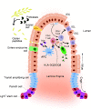
Figure 1.
Figure 1.
Schematic overview of celiac disease (CeD) pathobiology. Dietary gluten peptides pass the epithelial barrier, where they become deamidated by tissue transglutaminase 2 (TG2). The deamidated gluten peptides are taken up by antigen presenting cells (APCs) and are presented to CD4+ T cells, exclusively in the context of human leukocyte antigen (HLA)-DQ2 or HLA-DQ8. Upon gluten presentation, CD4+ T cells produce, among other things, interleukin (IL)-21 and interferon-gamma (IFN-γ). This leads to gluten-specific antibody production by B cells and, in concert with IL-15 production by intestinal epithelial cells (IECs), activation of intraepithelial lymphocytes (IELs), which attack the IECs, leading to villous atrophy.
Figure 2.
Figure 2.
Limitations of the intestinal organoid system. Intestinal organoids are inconsistent in size and shape, which introduces variability in the results (see left panel). The closed configuration makes it technically challenging to access the lumen (apical side) of the organoids. This limits studies into interactions between intestinal epithelial cells and micro-organisms (such as commensal microbes or pathogens), studies into transepithelial transport (e.g. fluorescein isothiocyanate-dextran translocation as a measure of intestinal permeability) and analysis of luminally secreted components (see middle panel). Intestinal organoids are cultured in a static three-dimensional system as they are embedded in an extracellular matrix, which does not reflect the dynamic environment of the human intestine (see right panel).
Figure 3.
Figure 3.
Schematic presentation of a microengineered intestine-on-chip. Intestine-on-chip systems often consist of a top microfluidic channel, resembling the gut lumen, and a bottom microfluidic channel, resembling the lamina propria and vasculature. The channels are separated by a porous membrane on which epithelial cells can be seeded and are flanked by vacuum chambers to simulate peristalsis-like movements. Unidirectional fluid flow through the microfluidic channels and contractions of the vacuum chambers simulate the physical microenvironment of the human intestine. The intestine-on-chip presented here is based on the design of Emulate Inc, Boston, MA, USA.
Figure 4.
Figure 4.
The steps from patient or healthy individual to human induced pluripotent stem cell (hiPSC)-derived intestinal organoids and intestinal barrier-on-chip. The large population and patient biobanks that have been constructed worldwide contain genomic data and stored biological material, which allow for the selection of patient and healthy control material based on genetic makeup. hiPSCs can be derived from stored kidney epithelial cells from urine or from erythroblasts from stored peripheral blood mononuclear cell fractions. These materials are obtained in a minimally invasive manner. The hiPSC lines can then be differentiated into human intestinal organoids (HIOs), which can subsequently be seeded on a microfluidic intestine-on-chip system to form an intestinal-barrier-on-chip. Specific genetic factors can be studied by genetic engineering of hiPSCs using CRISPR/Cas9 technology. For example, CeD-associated risk alleles can be reverted to protective alleles.
Figure 5.
Figure 5.
Research opportunities using a human induced pluripotent stem cell (hiPSC)-derived intestine-on-chip. (a) Functioning of the intestinal epithelial barrier in patients with celiac disease (CeD) can be assessed with the intestine-on-chip system by performing different assays: (1) Tight junctions and adherence junctions can be labeled and visualized on-chip using microscopy. (2) Barrier permeability can be assessed by measuring transepithelial passing of fluorescein isothiocyanate (FITC)-dextran complexes. (3) Barrier integrity can be tested by incorporating electrodes on-chip to measure transepithelial electrical resistance (TEER). (4) The passing of gluten peptides across the barrier and the direct effect of gluten peptides on the intestinal epithelial cells can be analyzed. (5) The effect of CeD-associated cytokines on the barrier can be analyzed by introducing the cytokines at the basolateral side (bottom channel). (b) Integration of gut microbiome, endothelial cells and immune cells in the intestine-on-chip. hiPSC-derived epithelial layers-on-chip can be extended with microbiomes from CeD patients or healthy controls on the apical side to assess the interactions between the epithelial layer and bacteria. hiPSC-derived endothelial cells can be introduced at the basolateral side to mimic the vascular system. Additionally, peripheral blood mononuclear cell (PBMC)- or hiPSC-derived immune cells can be introduced at the basolateral side to mimic the immune system.

References

    1. Biagi F, Klersy C, Balduzzi D, et al. Are we not over-estimating the prevalence of coeliac disease in the general population?. Ann Med 2010; 42: 557–561. - PubMed
    1. Bouziat R, Hinterleitner R, Brown JJ, et al. Reovirus infection triggers inflammatory responses to dietary antigens and development of celiac disease. Science 2017; 356: 44–50. - PMC - PubMed
    1. Harley JB, Chen X, Pujato M, et al. Transcription factors operate across disease loci, with EBNA2 implicated in autoimmunity. Nat Genet 2018; 50: 699–707. - PMC - PubMed
    1. Caminero A, Galipeau HJ, McCarville JL, et al. Duodenal bacteria from patients with celiac disease and healthy subjects distinctly affect gluten breakdown and immunogenicity. Gastroenterology 2016; 151: 670–683. - PubMed
    1. Spijkerman M, Tan IL, Kolkman JL, et al. A large variety of clinical features and concomitant disorders in celiac disease—A cohort study in the Netherlands. Dig Liver Dis 2016; 48: 499–505. - PubMed

Publication types

MeSH terms