. 2019 Apr;6(2):024003.
doi: 10.1117/1.JMI.6.2.024003. Epub 2019 Apr 29.

Three-dimensional conditional random field for the dermal-epidermal junction segmentation

Affiliations

Three-dimensional conditional random field for the dermal-epidermal junction segmentation

Julie Robic et al. J Med Imaging (Bellingham). 2019 Apr.

Abstract

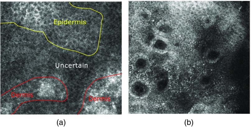
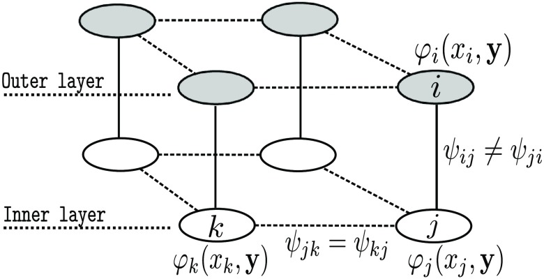
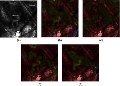
The segmentation of the dermal-epidermal junction (DEJ) in in vivo confocal images represents a challenging task due to uncertainty in visual labeling and complex dependencies between skin layers. We propose a method to segment the DEJ surface, which combines random forest classification with spatial regularization based on a three-dimensional conditional random field (CRF) to improve the classification robustness. The CRF regularization introduces spatial constraints consistent with skin anatomy and its biological behavior. We propose to specify the interaction potentials between pixels according to their depth and their relative position to each other to model skin biological properties. The proposed approach adds regularity to the classification by prohibiting inconsistent transitions between skin layers. As a result, it improves the sensitivity and specificity of the classification results.

Keywords: biomedical imaging; in vivo microscopy; machine learning; reflectance confocal microscopy.


Figures

Fig. 1
Fig. 1
Examples of different DEJ patterns. The circular rings pattern in (a) provides an easy identification of the DEJ compared to the uncertain one in (b). However, the latter one is the most frequent, especially on the cheeks.
Fig. 2
Fig. 2
Three-dimensional CRF modelization. The set of nodes in gray and in white belong to two different en-face sections. The edge potentials of each en-face sections ψjk [Eq. (3)] are learned at each depth. Edge potentials between en-face sections ψij [Eq. (3)] impose biological transition constraints.
Fig. 3
Fig. 3
Laplacian variance and distance to the closest minimum for a pixel between 20 and 120  μm. The blue line represents the Laplacian variance at coordinates (i,j) at all depths. The dashed vertical lines mark the position of the minimum of the Laplacian variance. The red line corresponds to the distance to the closest minimum.
Fig. 4
Fig. 4
Segmentations at depth d: epidermis (red), uncertain (yellow), dermis (blue). (a) Annotated image at depth d, (b) RF at depth d, (c) CRF2-D at depth d, (d) CRF3-DSym at depth d, and (e) CRF3-DAsym at depth d. The addition of constraints into the CRF model improves the accuracy of the segmentation.
Fig. 5
Fig. 5
Segmentations at depth d+1. (a) Annotated image at depth d+1, (b) RF at depth d+1, (c) CRF2-D at depth d+1, (d) CRF3-DSym at depth d+1, and (e) CRF3-DAsym at depth d+1. Inconsistent transitions exist between depth d and d+1. One can notice the misclassification obtained by RF and CRF2-D. The use of CRF3-DAsym provides a coherent segmentation.
Fig. 6
Fig. 6
Visual appearance of the transition from the epidermis to the uncertain area, which corresponds to the epidermal lower boundary. Notice that the aged DEJ (b) appears flatter than the young DEJ (a), as expected. Colors encode the depth.

References

    1. Lavker R. M., Zheng P., Dong G., “Aged skin: a study by light, transmission electron, and scanning electron microscopy,” J. invest. Dermatol. 88, 44s–51s (1987).JIDEAE10.1111/jid.1987.88.issue-s3 - DOI - PubMed
    1. Rittié L., Fisher G. J., “Natural and sun-induced aging of human skin,” Cold Spring Harbor Perspect. Med. 5(1), a015370 (2015).10.1101/cshperspect.a015370 - DOI - PMC - PubMed
    1. Calzavara-Pinton P., et al. , “Reflectance confocal microscopy for in vivo skin imaging,” Photochem. Photobiol. 84(6), 1421–1430 (2008).PHCBAP10.1111/php.2008.84.issue-6 - DOI - PubMed
    1. Pellacani G., et al. , “Reflectance confocal microscopy as a second-level examination in skin oncology improves diagnostic accuracy and saves unnecessary excisions: a longitudinal prospective study,” Br. J. Dermatol. 171(5), 1044–1051 (2014).BJDEAZ10.1111/bjd.2014.171.issue-5 - DOI - PubMed
    1. Robic J., et al. , “Automated quantification of the epidermal aging process using in-vivo confocal microscopy,” in IEEE 13th Int. Symp. Biomed. Imaging (ISBI), IEEE, pp. 1221–1224 (2016).10.1109/ISBI.2016.7493486 - DOI