. 2020 May 18;12(4):305-317.
doi: 10.1093/jmcb/mjz030.

Overexpression of mechanical sensitive miR-337-3p alleviates ectopic ossification in rat tendinopathy model via targeting IRS1 and Nox4 of tendon-derived stem cells

Affiliations

Overexpression of mechanical sensitive miR-337-3p alleviates ectopic ossification in rat tendinopathy model via targeting IRS1 and Nox4 of tendon-derived stem cells

Yiyun Geng et al. J Mol Cell Biol. .

Abstract

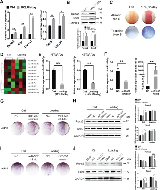
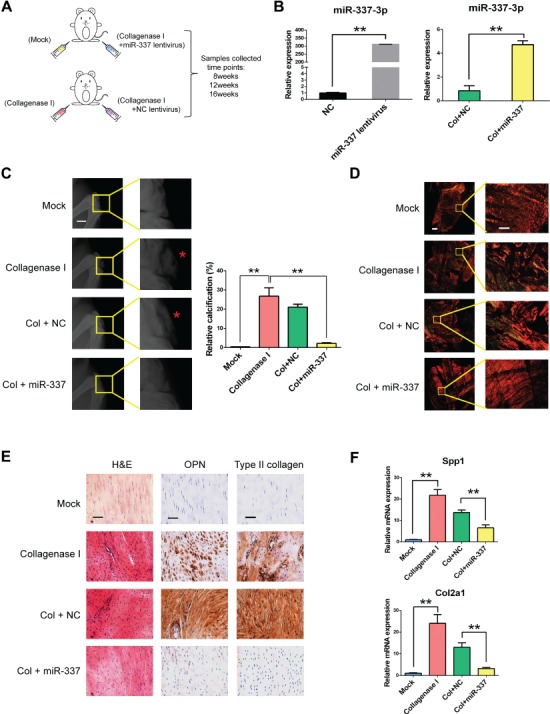
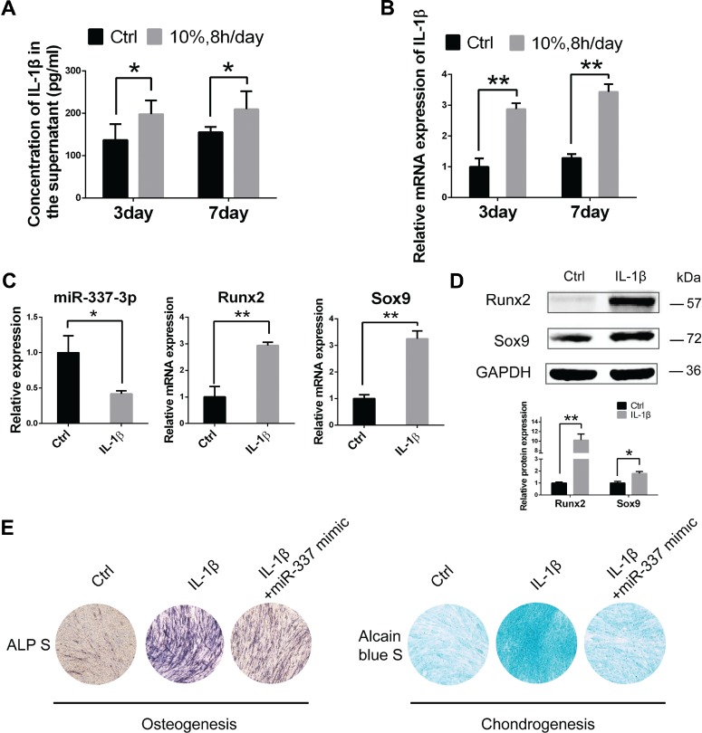
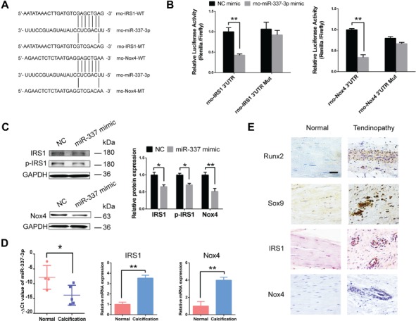
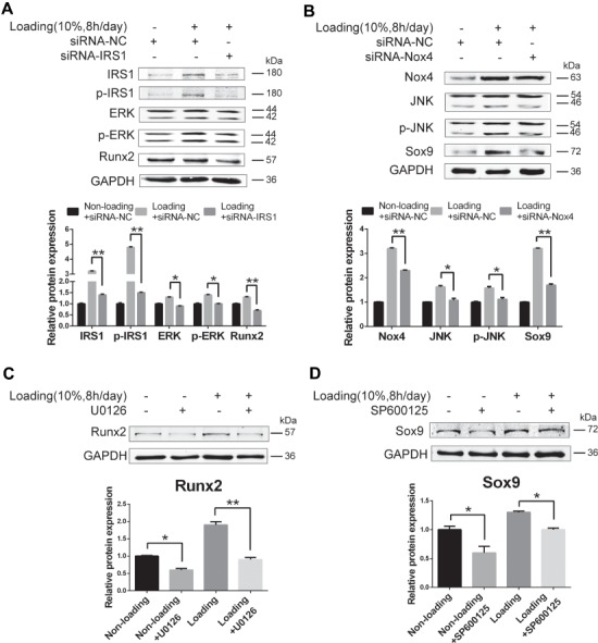
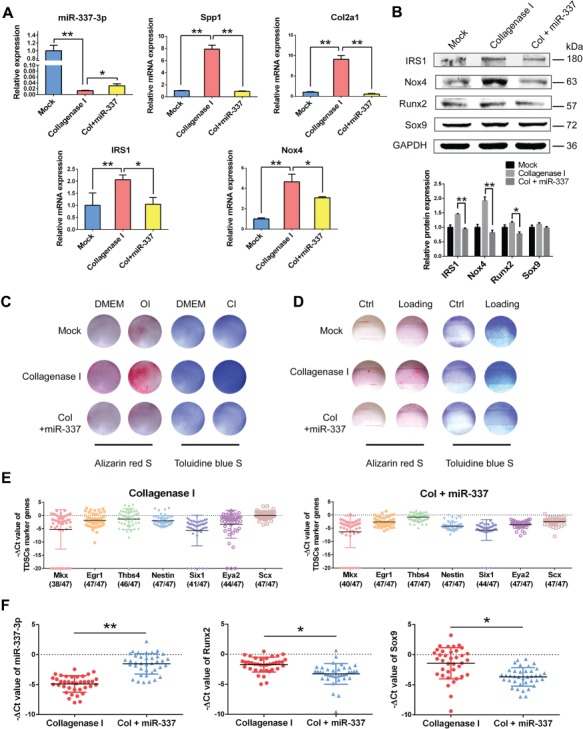
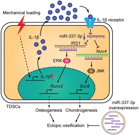
Tendinopathy, which is characterized by the ectopic ossification of tendon, is a common disease occurring in certain population, such as athletes that suffer from repetitive tendon strains. However, the molecular mechanism underlying the pathogenesis of tendinopathy caused by the overuse of tendon is still lacking. Here, we found that the mechanosensitive miRNA, miR-337-3p, had lower expression under uniaxial cyclical mechanical loading in tendon-derived stem cells (TDSCs) and negatively controlled chondro-osteogenic differentiation of TDSCs. Importantly, downregulation of miR-337-3p expression was also observed in both rat and human calcified tendons, and overexpressing miR-337-3p in patellar tendons of rat tendinopathy model displayed a robust therapeutic efficiency. Mechanistically, we found that the proinflammatory cytokine interleukin-1β was the upstream factor of miR-337-3p that bridges the mechanical loading with its downregulation. Furthermore, the target genes of miR-337-3p, NADPH oxidase 4, and insulin receptor substrate 1, activated chondro-osteogenic differentiation of TDSCs through JNK and ERK signaling, respectively. Thus, these findings not only provide novel insight into the molecular mechanisms underlying ectopic ossification in tendinopathy but also highlight the significance of miR-337-3p as a putative therapeutic target for clinic treatment of tendinopathy.

Keywords: chondro-osteogenesis; mechanical loading; mechanosensitive miRNA; tendinopathy; tendon-derived stem cells.


Figures

Figure 1
Figure 1
Mechanical loading promotes chondro-osteogenesis of TDSCs through downregulating miR-337-3p. (A) Real-time PCR analysis of osteogenic genes (left) and chondrogenic genes (right) in rTDSCs with or without mechanical loading (10%, 8 h/day) on Day 3. Ctrl stands for the non-loading group. Gapdh expression was used as an internal control for mRNA expression. (B) Western blot analysis of chondro-osteogenic genes in rTDSCs with or without mechanical loading (10%, 8 h/day) on Day 3. The densitometric analysis of Sox9 and Runx2 protein expression was normalized to GAPDH. Three independent experiments were analyzed for the bar graph below. (C) Alizarin red staining (upper) and Toluidine blue staining (lower) of rTDSCs cultured for 14 days. (D) miRNA microarray analysis of rTDSCs in normal culture and under mechanical loading (10%, 8 h/day) for 7 days. Three samples in each group were analyzed. Eleven differentially expressed miRNAs after normalized analysis are presented in the heat map. (E) Real-time PCR analysis of miR-337-3p expression under mechanical loading in rTDSCs (left) and hTDSCs (right) on Day 7. (F) Real-time PCR analysis of miR-337-3p expression in rTDSCs transfected with miR-337-3p inhibitor (left) or miR-337-3p mimic (right) on Day 2. (GJ) Alkaline phosphatase staining of rTDSCs treated with siRNA to inhibit rno-miR-337-3p (G) or rno-miR-337 mimics (I) along with mechanical loading (10%, 8 h/day) or not for 7 days. Protein levels of Runx2 and Sox9 of rTDSCs in rno-miR-337-3p inhibitor (H) or rno-miR-337 mimics treatment (J) combined with mechanical loading or not for 3 days. NC stands for negative control siRNA. Three independent experiments of western blot were analyzed for the bar graphs on the right. Mock stands for only transfection reagents-treated group. Error bars, SEM (n = 3). *P < 0.05; **P < 0.01.
Figure 2
Figure 2
miR-337-3p counteracts chondro-osteogenesis in collagenase I-induced rat tendinopathy model. (A) Schematic diagram of applying rno-miR-337-3p overexpressing lentivirus to cure rat tendinopathy induced by collagenase I. All reagents were injected at patellar tendon. Male SD rats (8-week-old) were used for the experiment, and patellar tendon samples were collected after 8, 12, and 16 weeks. Mock group stands for saline injection and suture. (B) Real-time PCR analysis of miR-337-3p in rTDSCs transfected with miR-337-3p overexpressing lentivirus for 7 days (left) and in patellar tendon tissues of rat tendinopathy model treated with miR-337-3p overexpressing lentivirus (right). (C) X-ray image of the knees of SD rats at 12 weeks after treatment showed that knee joints and ectopic ossicles (red asterisks in the magnified pictures) formed in patellar tendons (left). Scale bar, 5 mm. Relative calcification percents were measured through X-ray results by Image J (right). (D) Patellar tendon paraffin sections collected from each group at 12 weeks were treated with Sirius red staining and observed under polarized light microscopy to observe the collagen fibers. Left columns are images of integrated intact patellar tendon. Right columns are enlarged partial patellar tendon images. Scale bar, 800 μm (left) and 200 μm (right). (E) H&E staining and immunocytochemistry staining of OPN and type II collagen in each group. Samples were collected at 12 weeks after surgery. Scale bar, 50 μm. (F) Real-time PCR analysis of Spp1 and Col2a1 in patellar tendon tissues of each group. Error bars, SEM (n = 3). **P < 0.01.
Figure 3
Figure 3
Increased IL-1β induces miR-337-3p-mediated chondro-osteogenesis of rTDSCs under mechanical loading. (A) Enzyme-linked immunosorbent assay detected IL-1β in the supernatant medium of rTDSCs under mechanical loading (10%, 8 h/day) or not for 3 or 7 days. (B and C) Real-time PCR analysis of IL-1β (B), rno-miR-337-3p, Runx2, and Sox9 (C) in rTDSCs treated with 10 ng/ml IL-1β for 3 days. (D) Western blot analysis of Sox9 and Runx2 in rTDSCs treated with 10 ng/ml IL-1β for 3 days. The densitometric analysis of the proteins was normalized to GAPDH. Three independent experiments were analyzed for the bar graph below. (E) Alkaline phosphatase staining on Day 7 and Alcain blue staining on Day 14 of rTDSCs treated with IL-1β and rescued by rno-miR-337-3p mimics. Error bars, SEM (n = 3). *P < 0.05; **P < 0.01.
Figure 4
Figure 4
miR-337-3p targets IRS1 and Nox4 in tendinopathy. (A) Schematic representation of the rno-IRS1 and rno-Nox4 3′UTR indicating the binding sites of rno-miR-337-3p. WT, wild-type; MT, mutant. (B) HEK293T cells were transfected with psiCHECK™-2 Vector containing a fragment of rno-IRS1, rno-Nox4 3′UTR harboring binding sites for rno-miR-337-3p, or the corresponding mutant constructs. The effect of rno-miR-337 mimics on the corresponding vector luciferase activity was tested. (C) Protein levels of IRS1, p-IRS1, and Nox4 in rTDSCs treated with miR-337 mimics or negative control siRNAs (NC) for 3 days. The densitometric analysis of each protein expression was normalized to GAPDH. Three independent experiments were analyzed for the bar graph on the right. (D) Real-time PCR analysis of miR-337-3p, IRS1, and Nox4 expression in normal tendon tissues obtained from four osteoarthritis patients and calcified tendon tissues from five tendinopathy patients. (E) Immunocytochemistry staining of Runx2, Sox9, IRS1, and Nox4 in diseased tendon of tendinopathy patients and normal tendon samples. The densitometric analysis of the proteins was normalized to GAPDH. Error bars, SEM (n = 3). *P < 0.05; **P < 0.01. Scale bar, 50 μm.
Figure 5
Figure 5
ERK1/2 pathway activated by IRS1 and JNK pathway activated by Nox4 induce chondro-osteogenic differentiation of TDSCs under mechanical loading. (A and B) rTDSCs were transfected with IRS1 silencing RNAs (A), Nox4 silencing RNAs (B), or negative control siRNA combined with mechanical loading or not for 3 days. Western blot analysis of indicated proteins was presented. (C) ERK pathway inhibitor U0126 was applied to rTDSCs under mechanical loading condition or not for 3 days. Protein level of Runx2 was tested. (D) JNK pathway inhibitor SP600125 was applied to rTDSCs under mechanical loading condition or not for 3 days. Protein level of Sox9 was tested. The densitometric analysis of the proteins was normalized to GAPDH. The lower bands of ERK, p-ERK, JNK, and p-JNK were used for densitometric analysis. Three independent experiments were analyzed for the bar graph below. *P < 0.05; **P < 0.01. Scale bar, 50 μm.
Figure 6
Figure 6
rTDSCs from rat tendinopathy group and miR-337-3p curing group present different chondro-osteogenic differentiation ability. (A) Real-time PCR analysis of miR-337-3p and indicated genes of rTDSCs derived from each group. (B) Western blot analysis of miR-337-3p target genes and chondro-osteogenic genes in rTDSCs derived from each group. The densitometric analysis of each protein expression was normalized to GAPDH. Three independent experiments were analyzed for the bar graph below. (C) Alizarin red staining (left) and Toluidine blue staining (right) of rTDSCs derived from each group cultured in regular medium (DMEM), osteogenic-induced medium (OI), or chondrogenic-induced medium (CI) for 10 days. (D) Alizarin red staining (left) and Toluidine blue staining (right) of rTDSCs derived from each group with or without mechanical loading (10%, 8 h/day) for 10 days. (E and F) Single-cell PCR analysis of flow sorted rTDSCs (CD90+, CD45−) freshly derived from collagenase I-treated group and miR-337 overexpressing lentivirus group. (E) The expression of TDSC marker genes Mkx, Egr1, Thbs4, Nestin, Six1, Eya2, and Scx was normalized to Gapdh. (F) The expression of miR-337-3p, Runx2, and Sox9 in single cells with positive TDSC markers in either collagenase I-treated group or miR-337 overexpressing group. Numbers in parenthesis indicate cells positive for TDSC markers. Error bars, SEM (n = 3). *P < 0.05; **P < 0.01.
Figure 7
Figure 7
A schematic diagram shows that excessive mechanical loading-induced ectopic ossification is rescued by miR-337-3p overexpression. Downregulation of miR-337-3p by mechanical loading in TDSCs is mediated by the increase of inflammatory factor IL-1β. Nox4 and IRS1 are target genes of miR-337-3p. IRS1-activated JNK–Sox9 and Nox4-activated ERK–Runx2 pathways induce chondro-osteogenic differentiation of TDSCs, which contributes to the ectopic ossification of tendon.

References

    1. Ambros V. (2004). The functions of animal microRNAs. Nature 431, 350–355. - PubMed
    1. Aspenberg P. (2007). Is inflammation harmless to loaded tendons? J. Appl. Physiol. 102, 3–4. - PubMed
    1. Barwari T., Joshi A., and Mayr M. (2016). MicroRNAs in cardiovascular disease. J. Am. Coll. Cardiol. 68, 2577–2584. - PubMed
    1. Berkoff D.J., Kallianos S.A., Eskildsen S.M., et al. (2016). Use of an IL1-receptor antagonist to prevent the progression of tendinopathy in a rat model. J. Orthop. Res. 34, 616–622. - PubMed
    1. Bi Y., Ehirchiou D., Kilts T.M., et al. (2007). Identification of tendon stem/progenitor cells and the role of the extracellular matrix in their niche. Nat. Med. 13, 1219–1227. - PubMed

Publication types

MeSH terms