

Histologic Features Associated With an Invasive Component in Lentigo Maligna Lesions
- PMID: 31066867
- PMCID: PMC6506897
- DOI: 10.1001/jamadermatol.2019.0467
Histologic Features Associated With an Invasive Component in Lentigo Maligna Lesions
Abstract
Importance: Lentigo maligna (LM) presents an invasive component in up to 20% of biopsied cases, but to date the histologic features useful in detecting this invasive component have not been described. Some histologic characteristics are hypothesized to contribute to the progression of LM invasion.
Objective: To identify the histologic characteristics associated with lentigo maligna melanoma (LMM) in patients with LM diagnosed by a partial diagnostic biopsy.
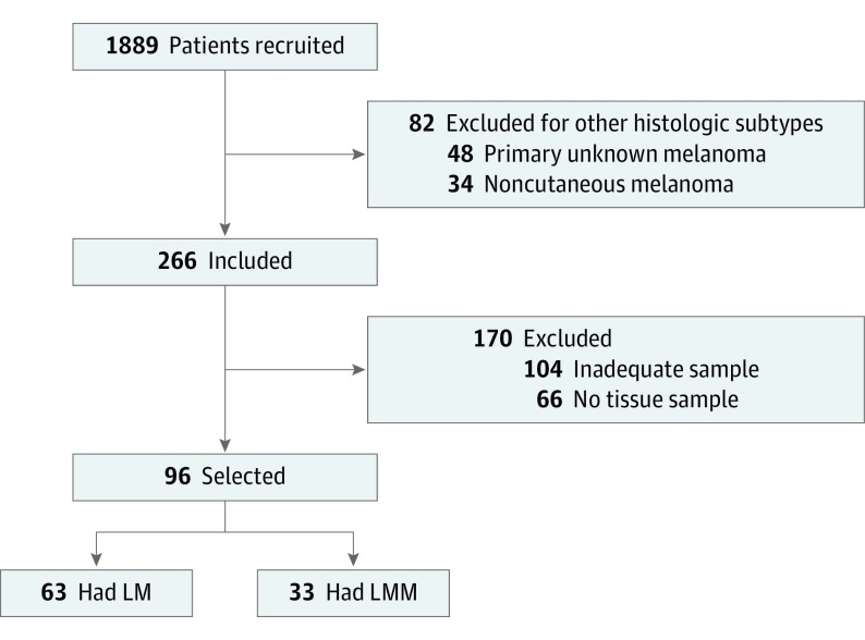
Design, setting, and participants: A retrospective cross-sectional study of patients treated between January 1, 2000, and December 31, 2017, was conducted in a referral oncology center in València, Spain. Data and specimens of patients (n = 96) with a diagnosis of primary cutaneous melanoma in the form of either LM or LMM who had undergone surgical treatment, a complete histologic examination of the whole tumor, and an initial diagnostic partial biopsy of LM were included in the study. Histologic assessment was blinded to the presence of an invasive component.
Interventions: All biopsy specimens were evaluated for the presence of certain histologic characteristics.
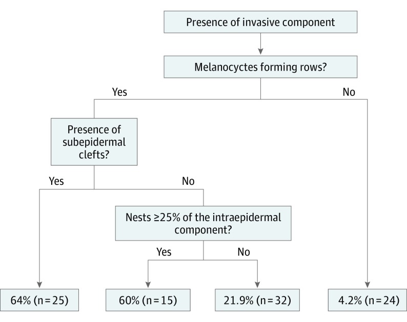
Main outcomes and measures: Comparisons between invasive samples and samples without an invasive component were performed. The differences in the distribution of variables between the groups were assessed using the χ2 and Fisher exact tests, and the degree of association of the relevant variables was quantified by logistic regression models. A classification and regression tree analysis was performed to rank the variables by importance.
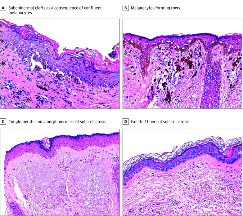
Results: In total, 96 patients had sufficient histologic material that could be evaluated. The patients were predominantly male (56 [58.3%]) and had a mean (SD) age at diagnosis of 72 (12) years. Of these patients, 63 (65.6%) had an LM diagnosis and 33 (34.4%) had an LMM diagnosis (an invasive component). The histologic variables associated with the presence of an invasive component were melanocytes forming rows (odds ratio [OR], 11.5; 95% CI, 1.4-94.1; P = .02), subepidermal clefts (OR, 2.8; 95% CI, 1.0-7.9; P = .049), nests (OR, 3.0; 95% CI, 1.1-8.6; P = .04), and a lesser degree of solar elastosis (OR, 0.4; 95% CI, 0.1-1.1; P = .07). A classification and regression tree analysis of the relevant histologic features was able to accurately identify lentigo maligna with an invasive component (LMM) in more than 60% of patients.
Conclusions and relevance: These findings may be useful in classifying early LM specimens at higher risk of invasion, which may eventually be relevant in identifying the most appropriate management for LM.
Conflict of interest statement
Figures
Comment in
-
The Morphology of Tumor Progression in Melanoma In Situ.JAMA Dermatol. 2019 Jul 1;155(7):775-776. doi: 10.1001/jamadermatol.2019.0457. JAMA Dermatol. 2019. PMID: 31066870 No abstract available.
References
-
- Elsner P, Diepgen TL, Schliemann S. Lentigo maligna and lentigo maligna melanoma as occupational skin diseases in a forestry worker with long-standing occupational UV-exposure. J Dtsch Dermatol Ges. 2014;12(10):915-917. - PubMed
MeSH terms
Grants and funding
LinkOut - more resources
Full Text Sources
Other Literature Sources
Medical
