. 2019 May 7;27(6):1755-1768.e4.
doi: 10.1016/j.celrep.2019.04.049.

Endothelial Cells in the Decidual Bed Are Potential Therapeutic Targets for Preterm Birth Prevention

Affiliations

Endothelial Cells in the Decidual Bed Are Potential Therapeutic Targets for Preterm Birth Prevention

Wenbo Deng et al. Cell Rep. .

Abstract

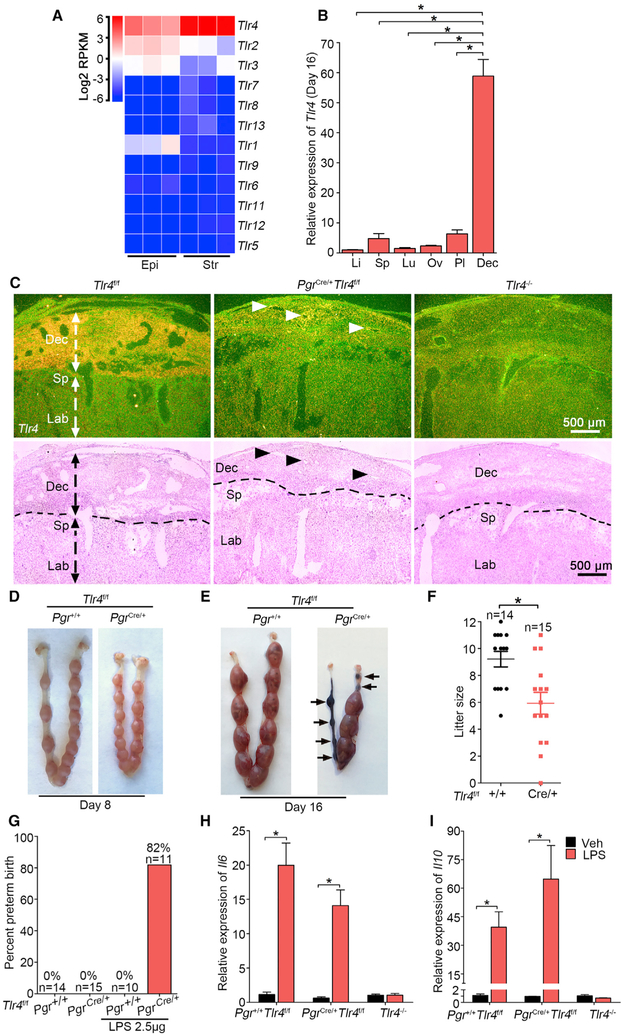
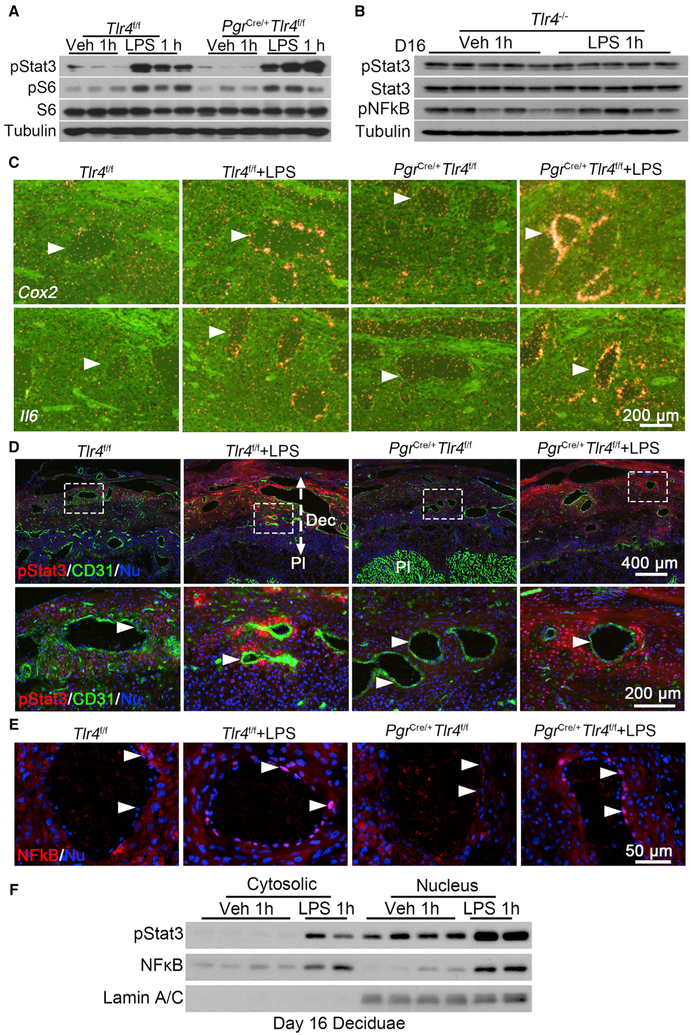
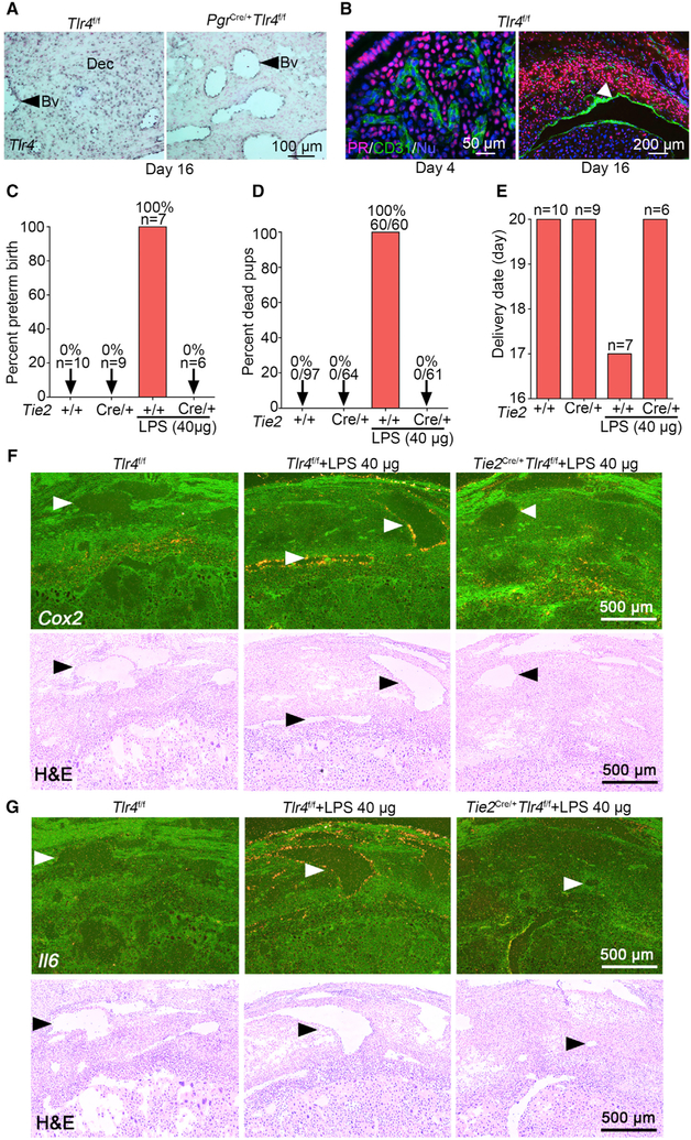
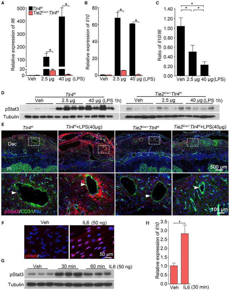
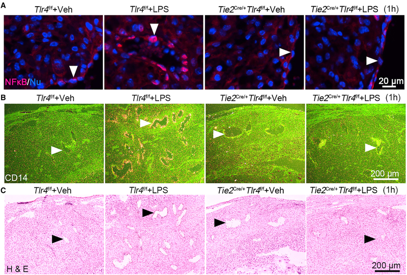
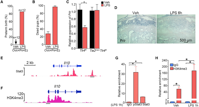
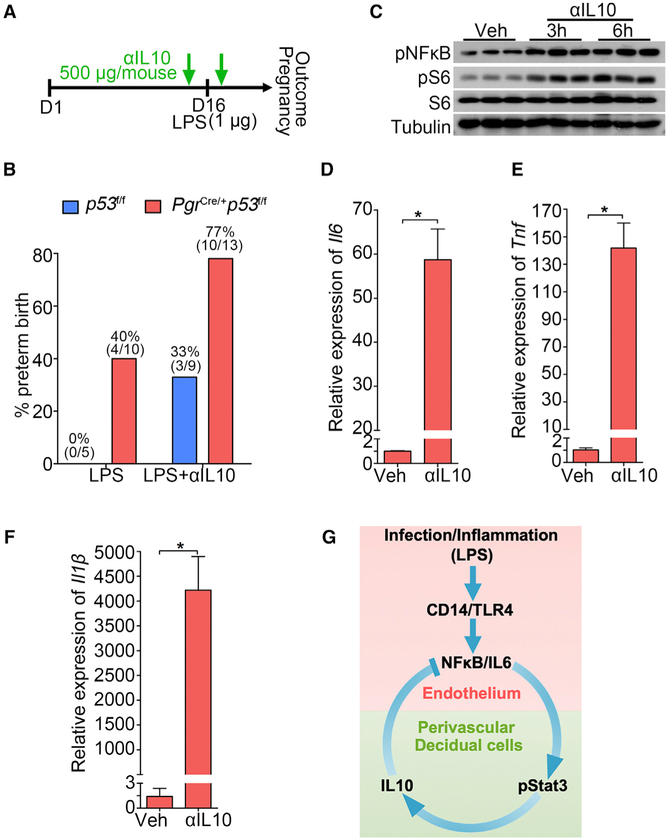
Preterm birth (PTB) is a syndrome with many origins. Among them, infection or inflammation are major risk factors for PTB; however, local defense mechanisms to mount anti-inflammatory responses against inflammation-induced PTB are poorly understood. Here, we show that endothelial TLR4 in the decidual bed is critical for sensing inflammation during pregnancy because mice with endothelial Tlr4 deletion are resistant to lipopolysaccharide (LPS)-induced PTB. Under inflammatory conditions, IL-6 is readily expressed in decidual endothelial cells with signal transducer and activator of transcription 3 (Stat3) phosphorylation in perivascular stromal cells, which then regulates expression of anti-inflammatory IL-10. Our observation that administration of an IL-10 neutralizing antibody predisposing mice to PTB shows IL-10's anti-inflammatory role to prevent PTB. We show that the integration of endothelial and perivascular stromal signaling can determine pregnancy outcomes. These findings highlight a role for endothelial TLR4 in inflammation-induced PTB and may offer a potential therapeutic target to prevent PTB.

Keywords: LPS; STAT3; TLR4; decidua; endothelial cell; pregnancy; preterm birth; uterus.


Conflict of interest statement

DECLARATION OF INTERESTS

The authors declare no competing interests.

Figures

Figure 1.
Figure 1.. TLR4 Is Beneficial during Normal Pregnancy
(A) Heatmap of RNA-seq analysis of Tlr mRNAs (log2 RPKM) in separated epithelial (Epi) and stromal cells (Str) from day 4 pregnant uteri (n = 3). RPKM, reads per kilobase per million. (B) Relative Tlr4 expression levels in various organs in day 16 pregnant Tlr4f/f mice by qPCR. rpL7 was used as internal controls. Li, liver; Sp, spleen; Lu, lung; Ov, ovary; Pl, placenta; Dec, decidua; n = 4, *p < 0.05. Data are represented as mean ± SEM. (C) Dark-field and bright-field images of in situ hybridization for Tlr4 in Tlr4f/f, PgrCre/+Tlr4f/f, and Tlr4 genomic knockout (Tlr4−/−) mice. Arrow heads indicate remaining Tlr4 in PgrCre/+Tlr4f/f mice. Dotted lines demarcate the interface between maternal decidua and spongiotrophoblast. Dec, decidua; Sp, spongiotrophoblast; Lb, labyrinth. Scale bar, 500 μm. (D and E) Days 8 (D) and 16 (E) implantation sites in Tlr4f/f and PgrCre/+Tlr4f/f mice. Arrows indicate resorption sites. (F) Live litter size in Tlr4f/f and PgrCre/+Tlr4f/f mice. n, animal number in each group, *p < 0.05. Data are represented as mean ± SEM. (G) The ratio of preterm birth to total number of pups per gestation in Tlr4f/f and PgrCre/+Tlr4f/f mice with or without exposure to a low dose LPS (2.5 μg/mouse). n indicates the number of total mice examined in each group. (H and I) qPCR of Il6 (H) and Il10 (I) mRNA levels in Tlr4f/f, PgrCre/+Tlr4f/f, and Tlr4−/− mice after 1 h of LPS treatment. *p < 0.05. Data are represented as mean ± SEM (n = 4). See also Figure S1.
Figure 2.
Figure 2.. Interactions between Endothelial and Perivascular Stromal Cells in the Decidual Bed of PgrCre/+Tlr4f/f Mice
(A) Western blotting of pStat3 and pS6 levels in day 16 uteri of Tlr4f/f and PgrCre/+Tlr4f/f dams exposed to LPS (2.5 μg/mouse), n = 3. (B) Western blotting results of pStat3, Stat3, and pNFkB levels in day 16 deciduae after LPS treatment for 1 hour in Tlr4−/− mice, n = 5. (C) In situ hybridization for Cox2 and Il6 mRNAs in Tlr4f/f and PgrCre/+Tlr4f/f mice after exposure to LPS for 1 hour. Arrowheads indicated endothelial cells. Scale bar, 200 μm. (D) Immunofluorescence staining of pStat3 (red) and CD31 (green) in Tlr4f/f and PgrCre/+Tlr4f/f mice after exposure to LPS for 1 hour. Scale bar of upper panels, 400 μm. Bottom panels show images at higher magnification of those within the demarcated rectangles in the upper panels. Dec, decidua; Pl, placenta. Arrowheads indicate endothelial cells. Scale bar, 200 μm. (E) Immunostaining showing nuclear NF-kB (red) in Tlr4f/f and PgrCre/+Tlr4f/f mice after exposure to LPS for 1 hour. Arrowheads indicate endothelial cells. Scale bar, 50 μm. (F) Western blotting for pStat3 and NF-kB in cytosolic and nuclear fractions of day 16 deciduae after 1 h of vehicle or LPS treatment in Tlr4f/f females. Lamin AC was used as nuclear internal control. See also Figures S2, S3, and S4.
Figure 3.
Figure 3.. Endothelial-Specific Tlr4 Deletion with a Tie2-Cre Driver Protects against LPS-Induced Preterm Birth
(A) DIG-in situ hybridization for Tlr4 in decidual endothelia of Tlr4f/f and PgrCre/+Tlr4f/f mice on day 16 of pregnancy. Blue color indicates the Tlr4 signal. Dec, decidua. Arrow heads indicate blood vessels (Bv). Scale bar, 100 μm. (B) PR (progesterone receptor)-positive cells are absent in the endothelium in days 4 (left) and 16 (right) uteri. Arrow head indicates blood vessel. Scale bars for the left and right panels represent 50 μm and 200 μm, respectively. (C and D) The ratio of preterm birth (C) and dead pups (D) compared to total number of pups in Tlr4f/f and Tie2Cre/+Tlr4f/f mice with or without a high dose of LPS (40 μg/mouse). n, number of dams examined. (E) Parturition timing in Tlr4f/f and Tie2Cre/+Tlr4f/f mice with or without LPS. n, number of dams examined. (F) Dark-field and bright-field images of in situ hybridization for Cox2 in Tlr4f/f and Tie2Cre/+Tlr4f/f deciduae after 1 h of LPS exposure (40 μg/mouse). Arrow heads indicate blood vessels. Scale bar, 500 μm. (G) Dark-field and bright-field images of in situ hybridization for Il6 in Tlr4f/f and Tie2Cre/+Tlr4f/f deciduae after 1 h of LPS exposure (40 μg/mouse). Arrow heads indicate blood vessels. Scale bar, 500 μm. See also Figure S4.
Figure 4.
Figure 4.. Endothelial TLR4 Is Critical for Sensing LPS-Induced Responses in Decidua
(A and B) Relative expression of Il6 (A) and Il10 (B) mRNAs in Tlr4f/f and Tie2Cre/+Tlr4f/f mice after a low dose (2.5 μg/mouse) or high dose (40 μg/mouse) of LPS treatment for 1 hour. n = 4. *p < 0.05. Results are shown as mean ± SEM. (C) Ratio of mRNA levels of Il10 versus Il6 in deciduae of Tlr4f/f mice under a low and high dose of LPS. n = 4. *p < 0.05. Results are shown as mean ± SEM. (D) western blotting of protein levels for pStat3 in Tlr4f/f and Tie2Cre/+Tlr4f/f mice after 1 h of a low or high dose of LPS treatment on day 16 of pregnancy, n = 4. (E) Co-immunostaining of pStat3 (red) and CD31 (green) in LPS-treated deciduae of Tlr4f/f and Tie2Cre/+Tlr4f/f mice. Scale bar of upper panels, 500 μm. Bottom panels show images of higher magnification of those within the demarcated rectangles (Scale bar, 100 μm). Arrowheads indicate endothelial cells. Dec, deciduae; Pl, placenta. (F) pStat3 nuclear translocation by IL-6. Isolated stromal cells were treated with recombinant IL-6 (50 ng) for 30 min. Scale bar, 50 μm. (G and H) pStat3 levels (G) and Il10 mRNA levels (H) after recombinant interleukin 6 (rIL-6) challenge. Tubulin and rpL7 were used as internal controls for western blotting and qPCR, respectively. n = 3. *p < 0.05. Results are shown as mean ± SEM. See also Figures S5, S6, and S7.
Figure 5.
Figure 5.. Endothelial NF-kB and CD14 Are Downregulated in Tie2Cre/+Tlr4f/f Deciduae
(A) Immunostaining shows nuclear translocation of NF-kB (red) in Tlr4f/f and Tie2Cre/+Tlr4f/f mice after exposure to LPS (40 μg/mouse) for 1 hour. Arrow heads indicate endothelial cells. Scale bar, 20 μm. (B and C) LPS-induced expression of Cd14 by radioactive in situ hybridization (B) in day 16 decidual endothelial cells in Tlr4f/f and Tie2Cre/+Tlr4f/f mice. The lower panel (C) represents H&E-stained images. Arrow heads indicate endothelial cells. Scale bar, 200 μm.
Figure 6.
Figure 6.. LPS Targets Deciduae, Not Ovaries, and Regulation of IL-10 by Stat3
(A) Incidence of PTB in pregnant mice ovariectomized on day 16 with or without exposure to LPS (40 μg/mouse) followed by P4 injections (1 mg) on days 16 and 17 and 250 μg of P4 on day 18 with an additional injection of estradiol-17β (100 ng) on day 19. All ovariectomized (Ovx) mice given P4 and E2 without LPS showed normal delivery, albeit 40% dead pups. In contrast, over 90% of LPS-treated mice delivered dead pups before day 19 with 100% dead pups. Ovx, ovariectomy. n, number of mice examined in each group. (B) Rate of dead pups in mice with or without LPS injection after ovariectomy followed by P4 and E2 injection. n = 12. (C) Relative expression of Prlr mRNA in Tlr4f/f and Tie2Cre/+Tlr4f/f mice after exposure to LPS (40 μg/mouse) for 1 hour. n = 4, *p < 0.05. Results are shown as mean ± SEM. (D) DIG-in situ hybridization for Prlr in Tlr4f/f mice after 6 h of LPS (40 μg/mouse) treatment. Scale bar, 500 μm. (E and G) Graphic representation of predicted Stat3 binding site on the Il10 gene (E) and ChIP-qPCR confirmation of binding by both pStat3 and Stat3 antibodies in deciduae after LPS injection for 1 h (G). n = 3, *p < 0.05. Data are represented as mean ± SEM. (F and H) Graphic representation of H3K4me3 enrichment at the Il10 promoter region (F) with ChIP-qPCR confirmation in deciduae after LPS injection for 1 h (H). n = 3, *p < 0.05. Data are represented as mean ± SEM.
Figure 7.
Figure 7.. The Role of IL-10 in PTB Prevention
(A) A scheme of treatment schedule of IL-10 neutralizing antibody (αIL-10). (B) Preterm birth rate of p53f/f and PgrCre/+p53f/f mice after IL-10 neutralizing antibody (αIL-10) injection in the presence of a ultra-low dose of LPS exposure (1 μg/mouse). The number in parentheses indicates the incidence of PTB in total number of mice analyzed. (C) Western blot results for pNFkB and pS6 levels in decidualized stromal cells after IL-10 neutralizing antibody treatment for 3 and 6 hours. (D–F) Il6 (D), Il1b (E), and Tnf (F) mRNA levels by IL-10 neutralizing antibody treatment. n = 3, *p < 0.05. Data are represented as mean ± SEM (n = 3). (G) A representative scheme of a physiological role of endothelial TLR4 in LPS-induced inflammation. After binding with LPS, endothelial TLR4 directs NFkB nuclear translocation to stimulate endothelial IL-6 expression in endothelial cells. Locally generated IL-6 stimulates Stat3 phosphorylation in perivascular decidual cells for IL-10 expression. Activation of endothelial TLR4 by LPS, which counters endogenous IL-10 production, results in preterm birth. CD14 is a co-receptor for TLR4 and highly induced in endothelial cells after LPS stimulation.

References

    1. Aisemberg J, Vercelli CA, Bariani MV, Billi SC, Wolfson ML, and Franchi AM (2013). Progesterone is essential for protecting against LPS-induced pregnancy loss. LIF as a potential mediator of the anti-inflammatory effect of progesterone. PLoS One 8, e56161. - PMC - PubMed
    1. Altmäe S, Koel M, Vôsa U, Adler P, Suhorut senko M, Laisk-Podar T, Kukushkina V, Saare M, Velthut-Meikas A, Krjut skov K, et al. (2017). Meta-signature of human endometrial receptivity: a meta-analysis and validation study of transcriptomic biomarkers. Sci. Rep 7, 10077. - PMC - PubMed
    1. Cha J, Bartos A, Egashira M, Haraguchi H, Saito-Fujita T, Leishman E, Bradshaw H, Dey SK, and Hirota Y (2013). Combinatory approaches prevent preterm birth profoundly exacerbated by gene-environment interactions. J. Clin. Invest 123, 4063–4075. - PMC - PubMed
    1. Choukrallah MA, Song S, Rolink AG, Burger L, and Matthias P (2015). Enhancer repertoires are reshaped independently of early priming and heterochromatin dynamics during B cell differentiation. Nat. Commun 6, 8324. - PMC - PubMed
    1. Clemente-Casares X, Blanco J, Ambalavanan P, Yamanouchi J, Singha S, Fandos C, Tsai S, Wang J, Garabatos N, Izquierdo C, et al. (2016). Expanding antigen-specific regulatory networks to treat autoimmunity. Nature 530, 434–440. - PubMed

Publication types

MeSH terms