. 2019 May 7;6(2):39.
doi: 10.3390/bioengineering6020039.

Elastin-Dependent Aortic Heart Valve Leaflet Curvature Changes During Cyclic Flexure

Affiliations

Elastin-Dependent Aortic Heart Valve Leaflet Curvature Changes During Cyclic Flexure

Melake D Tesfamariam et al. Bioengineering (Basel). .

Abstract

The progression of calcific aortic valve disease (CAVD) is characterized by extracellular matrix (ECM) remodeling, leading to structural abnormalities and improper valve function. The focus of the present study was to relate aortic valve leaflet axial curvature changes as a function of elastin degradation, which has been associated with CAVD. Circumferential rectangular strips (L × W = 10 × 2.5 mm) of normal and elastin-degraded (via enzymatic digestion) porcine AV leaflets were subjected to cyclic flexure (1 Hz). A significant increase in mean curvature (p < 0.05) was found in elastin-degraded leaflet specimens in comparison to un-degraded controls at both the semi-constrained (50% of maximum flexed state during specimen bending and straightening events) and fully-constrained (maximally-flexed) states. This significance did not occur in all three flexed configurations when measurements were performed using either minimum or maximum curvature. Moreover, the mean curvature increase in the elastin-degraded leaflets was most pronounced at the instance of maximum flexure, compared to un-degraded controls. We conclude that the mean axial curvature metric can detect distinct spatial changes in aortic valve ECM arising from the loss in bulk content and/or structure of elastin, particularly when there is a high degree of tissue bending. Therefore, the instance of maximum leaflet flexure during the cardiac cycle could be targeted for mean curvature measurements and serve as a potential biomarker for elastin degradation in early CAVD remodeling.

Keywords: aortic valve; biomarker; calcification; curvature; early detection; elastin degradation; leaflet.


Conflict of interest statement

The authors declare no conflict of interest.

Figures

Figure 1
Figure 1
Leaflet strip set-up in mechanical test instrument (Electroforce 2300 with Wintest 7 software control, TA instruments) to facilitate specimen cyclic flexure (bending and straightening events).
Figure 2
Figure 2
Workflow (A) Tissue preparation: i) Porcine aortic valve, ii) split aortic valve revealing the three leaflets, iii) schematic of circumferentially oriented rectangular strips cut from the belly region of porcine aortic valve leaflets. The remaining surrounding tissue was minced and used for the degradation and quantification of elastin in this study. (B) Image processing: i) Cropped image according to region of interest, ii) increase background to foreground contrast, iii) threshold image, iv) edge detection. (C) Curvature quantification: i) Left/center/right curvatures for a frame of a video, ii) temporal curvature for a given leaflet, iii) spatial curvature for a given leaflet, iv) minimum, Maximum, and mean curvature along center profile for the 5–10 range.
Figure 3
Figure 3
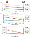
Elastin and curvature comparisons. (A) Maximum curvature with mean ± SEM plotted against elastin for 0 h and 2 h degradation time points for all bending phases. (B) Mean curvature with mean ± SEM plotted against elastin for 0 h and 2 h degradation time points for all bending phases. (C) Minimum curvature with mean ± SEM plotted against elastin for 0 h and 2 h degradation time points for all bending phases.
Figure 4
Figure 4
(AD) Elastin structure in aortic valve leaflets before (A,C) and after (B,D) elastase-degradation. Images (A) and (B) were obtained from the non-coronary cusp whereas images (C) and (D) were from the left coronary cusp. Image sections were taken at a depth of 36 µm from the nearest leaflet surface. Scale bar in all images is 50 µm. Elastin fibers (A,C) exhibited a strong affinity for alignment in the radial direction. Enzymatic-degradation clearly led to loss of elastin content, orientation and network in the leaflets (B,D). (EH) Collagen distribution at the same spatial location in which Elastin structure was imaged (AD). Collagen fibers (E,G) were primarily oriented in circumferential direction. Degradation with elastase enzyme was not specific to elastin as clearly, loss of collagen content, orientation and network in the leaflets also occurred (F,H).
Figure 5
Figure 5
Movat’s staining of: (A) Normal native porcine aortic valve non-coronary leaflet and (B) elastase-degraded (0.5 mg/mL for 2 h) native porcine aortic valve non-coronary leaflet. The extracellular matrix (ECM) components stain as brownish-black for Elastin and yellow-orange for Collagen. “F” indicates the fibrosa side and “V” is the ventricularis side of the leaflet. Note that the ventricularis side of the leaflet was completely lost after elastase degradation (B).

References

    1. Perrotta I., Camastra C., Filice G., Di Mizio G., Colosimo F., Ricci P., Tripepi S., Amorosi A., Triumbari F., Donato G., et al. New evidence for a critical role of elastin in calcification of native heart valves: Immunohistochemical and ultrastructural study with literature review. Histopathology. 2011;59:504–513. doi: 10.1111/j.1365-2559.2011.03977.x. - DOI - PubMed
    1. Passik C.S., Ackermann D.M., Pluth J.R., Edwards W.D. Temporal changes in the causes of aortic stenosis: A surgical pathologic study of 646 cases. Mayo Clin. Proc. 1987;62:119–123. doi: 10.1016/S0025-6196(12)61880-1. - DOI - PubMed
    1. Rutkovskiy A., Malashicheva A., Sullivan G., Bogdanova M., Kostareva A., Stensløkken K., Fiane A., Vaage J. Valve interstitial cells: The key to understanding the pathophysiology of heart valve calcification. J. Am. Heart Assoc. 2017;6:6. doi: 10.1161/JAHA.117.006339. - DOI - PMC - PubMed
    1. Yutzey K.E., Demer L.L., Body S.C., Huggins G.S., Towler D.A., Giachelli C.M., Hofmann-Bowman M.A., Mortlock D.P., Rogers M.B., Sadeghi M.M., et al. Calcific aortic valve disease: A consensus summary from the Alliance of Investigators on Calcific Aortic Valve Disease. Arterioscler. Thromb. Vasc. Biol. 2014;34:2387–2393. doi: 10.1161/ATVBAHA.114.302523. - DOI - PMC - PubMed
    1. Engelmayr G.C., Sales V.L., Mayer J.E., Sacks M.S. Cyclic flexure and laminar flow synergistically accelerate mesenchymal stem cell-mediated engineered tissue formation: Implications for engineered heart valve tissues. Biomaterials. 2006;27:6083–6095. doi: 10.1016/j.biomaterials.2006.07.045. - DOI - PubMed

LinkOut - more resources