

Adrenal hemorrhage in newborn: how, when and why- from case report to literature review
- PMID: 31068206
- PMCID: PMC6507044
- DOI: 10.1186/s13052-019-0651-9
Adrenal hemorrhage in newborn: how, when and why- from case report to literature review
Abstract
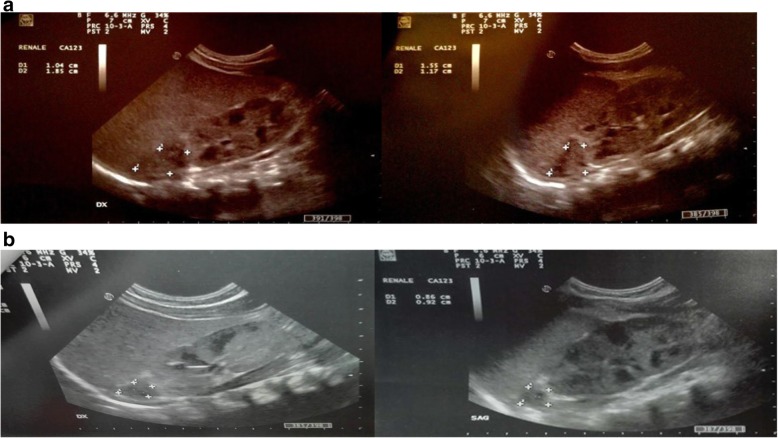
Background: Neonatal adrenal hemorrhage is a relatively uncommon condition (0.2-0.55%). Various risk factors have been reported in addition to birth asphyxia, such as sepsis, coagulation disorders, traumatic delivery, and perinatal injuries. Adrenal hemorrhage usually affects the right adrenal gland (about 70% of cases) while it involves the bilateral adrenal gland only in 10% of cases. In most cases, the event is asymptomatic but, in others, it may be so devastating to determine death by bleeding or adrenal insufficiency.
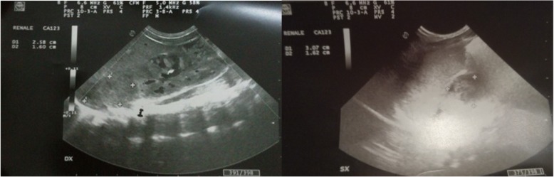
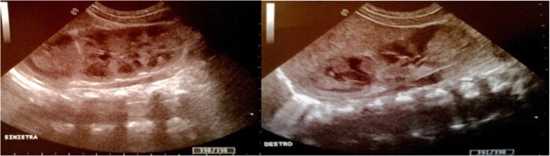
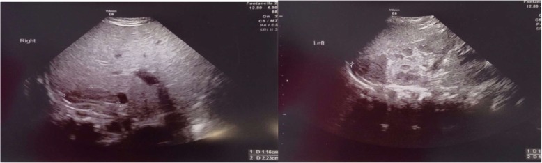
Case presentation: A case of bilateral neonatal adrenal hemorrhage, with adrenal insufficiency, but with no important risk factors and favorable evolution in a male infant.
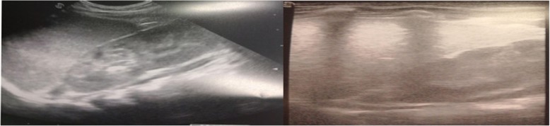
Conclusions: This case emphasizes the importance of keeping a non-interventional attitude, avoiding early surgery but carrying out a serial sonographic follow-up. Serial ultrasound monitoring is the most reliable approach during conservative management.
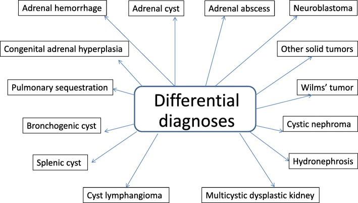
Keywords: Adrenal insufficiency; Differential diagnosis; Hormonal therapy; Neonatal adrenal hemorrhage; Ultrasound monitoring.
Conflict of interest statement
Ethics approval and consent to participate
Not applicable.
Consent for publication
The patient’s parent provides his consent to submission.
Competing interests
The authors declare that they have no competing interests.
Publisher’s Note
Springer Nature remains neutral with regard to jurisdictional claims in published maps and institutional affiliations.
Figures
References
-
- Bergami G, Malena S, Di Mario M, Fariello G. Ecography in the follow-up of neonatal AH. The presentation of 14 cases. Radiol Med. 1990;79(5):474–478. - PubMed
-
- Lee MC, Lin LH. Ultrasound screening of neonatal adrenal hemorrhage. Acta Paediatr Taiwan. 2000;41(6):327–330. - PubMed
-
- Wang CH, Chen AJ, Yang LY, Tang R.B. Neonatal adrenal hemorrhage presenting as a multiloculated cystic mass. J Chin Med Assoc 2008; vol 71, n 9. - PubMed
Publication types
MeSH terms
LinkOut - more resources
Full Text Sources
Medical
