. 2019 May 8;10(1):2098.
doi: 10.1038/s41467-019-10117-z.

Enveloped viruses distinct from HBV induce dissemination of hepatitis D virus in vivo

Affiliations

Enveloped viruses distinct from HBV induce dissemination of hepatitis D virus in vivo

Jimena Perez-Vargas et al. Nat Commun. .

Abstract

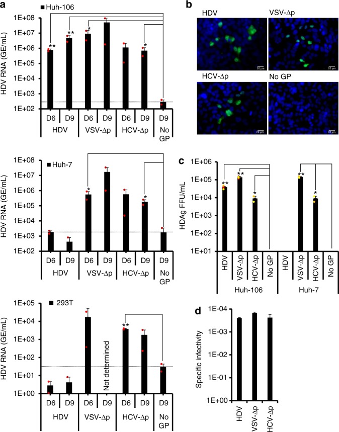
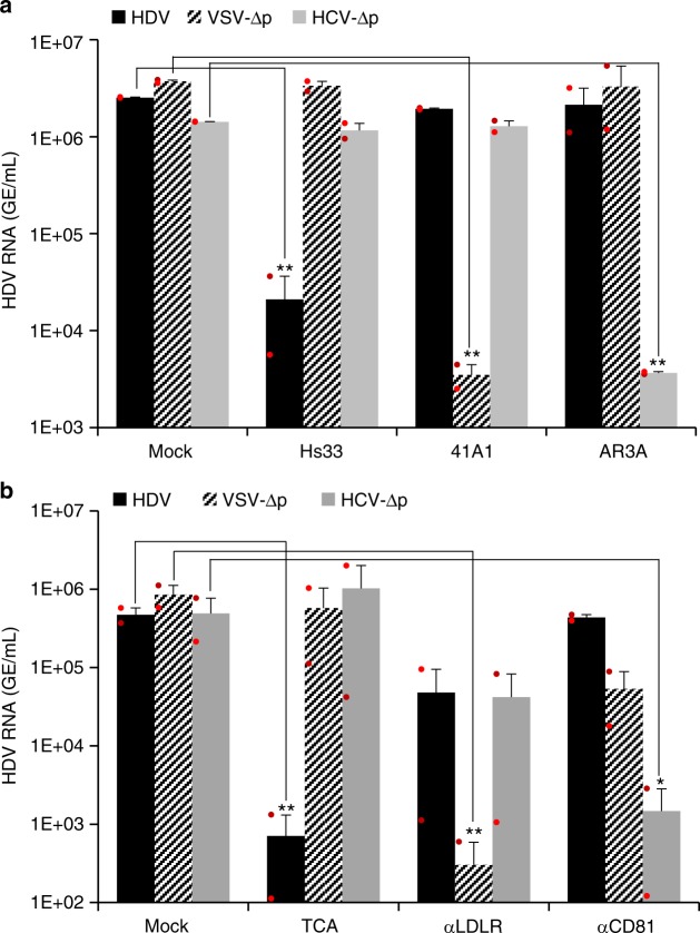
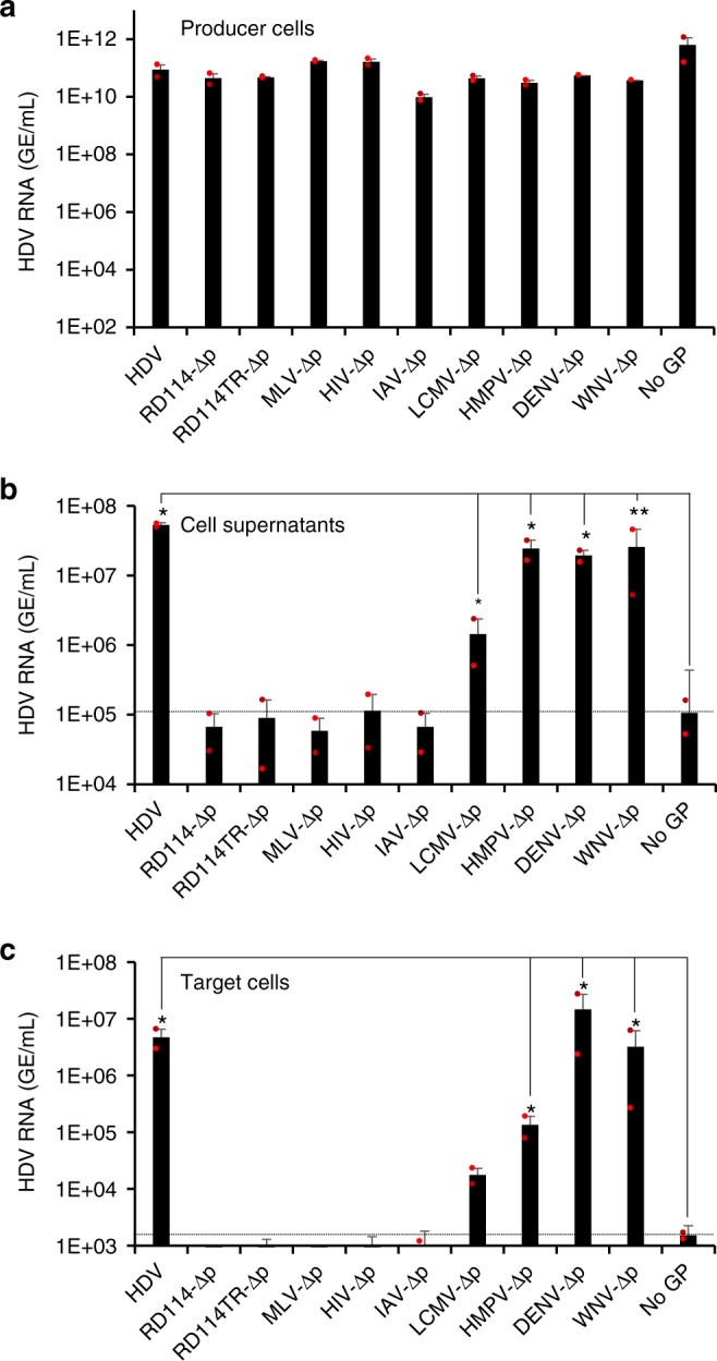
Hepatitis D virus (HDV) doesn't encode envelope proteins for packaging of its ribonucleoprotein (RNP) and typically relies on the surface glycoproteins (GPs) from hepatitis B virus (HBV) for virion assembly, envelopment and cellular transmission. HDV RNA genome can efficiently replicate in different tissues and species, raising the possibility that it evolved, and/or is still able to transmit, independently of HBV. Here we show that alternative, HBV-unrelated viruses can act as helper viruses for HDV. In vitro, envelope GPs from several virus genera, including vesiculovirus, flavivirus and hepacivirus, can package HDV RNPs, allowing efficient egress of HDV particles in the extracellular milieu of co-infected cells and subsequent entry into cells expressing the relevant receptors. Furthermore, HCV can propagate HDV infection in the liver of co-infected humanized mice for several months. Further work is necessary to evaluate whether HDV is currently transmitted by HBV-unrelated viruses in humans.


Conflict of interest statement

The authors declare no competing interests.

Figures

Fig. 1
Fig. 1
Secretion of HDV particles is induced by surface glycoproteins from varied enveloped viruses. Huh-7 cells were co-transfected with pSVLD3 plasmid coding for HDV RNPs and plasmids coding for HBV, VSV, or HCV surface glycoproteins (GP), resulting in “HDV”, “VSV-∆p”, and “HCV-∆p” samples, respectively. As control, pSVLD3 was co-transfected with an empty plasmid (“No GP” samples). a At day 3, 6, or 9, extracellular HDV RNAs were quantified from cell supernatants by RT-qPCR. Intracellular HDV RNAs were quantified from cell lysates at day 9 post transfection. HDV RNA levels in GE (genome equivalent) are expressed as means (n = 5 independent experiments) per ml of cell supernatants for extracellular RNAs or, for intracellular RNAs, per mL of cell lysates containing 106 cells. b RNAs extracted from lysates and supernatants of transfected cells treated with RNAse-free DNAse, or not treated (–DNAse), were reverse-transcribed using a antigenomic primer that detects HDV RNAs and then PCR-amplified with HDV-specific primers to reveal unit-length HDV genomic RNAs. As control, reverse transcriptase was omitted during processing of the samples (–RT). c, d In total, 2 × 107 HDV GEs from pellets retrieved after ultracentrifugation of cell supernatants on 30% sucrose cushions were analyzed by northern blot using a HDV-specific probe (c) or by western blot using an HDAg antibody (d). Control HDV RNAs (5 × 107 GE) (c) or HDAg from cell lysates (d) were loaded on the same gels (Ctrl). e Pelleted cell supernatants containing 109 HDV GEs (“Input”) immunoprecipitated with antibodies against HBsAg (Hs33 mAb), VSV-G (41A1 mAb), and HCV-E1E2 (AR3A mAb) glycoproteins, as indicated, were quantified by RT-qPCR after elution. The results are expressed as percentages of input values. f Electron microscopy of heparin bead-purified supernatants after elution and negative staining showing large (white arrows) and small (black arrows) particles. Scale bar: 100 nm. g HDV RNAs, from fractions from cell supernatant samples separated on equilibrium-density gradients, were analyzed by RT-qPCR, expressed as percentages of total HDV RNA contents, or by strand-specific RT-PCR that reveals HDV genome size (below each graph). Source data are provided as a Source Data file. Error bars correspond to standard deviation. Statistical analyses (Student’s t-test): p < 0.05 (*)
Fig. 2
Fig. 2
HDV particles generated with heterologous envelope glycoproteins are infectious. a The infectivity of virus particles produced with HBV (HDV), VSV (VSV-∆p), HCV (HCV-∆p) glycoproteins, or with no envelope glycoprotein (No GP) and harvested at day 6 or 9 post transfection (see Fig. 1a) was determined in Huh-106 (NTCP-expressing Huh-7 cells), Huh-7, or 293T cells, as indicated. Infected cells were grown for 7 days before total intracellular RNA was purified. The results of HDV RNA quantification by RT-qPCR are expressed as means (n = 2 independent experiments) per mL of cell lysates containing 105 cells. Nd, not determined. The dotted lines represent the experimental thresholds, as defined with the “No GP” controls. b, c Huh-106 and Huh-7 cells infected by serial dilutions of supernatants containing the indicated virus particles harvested at day 9 post transfection (Fig. 1a) were fixed at 7 days post infection and stained by immunofluorescence with the SE1679 polyclonal anti-HDAg antibody before counting the foci of HDAg-positive cell colonies. The cells were counterstained with Hoechst to visualize the nuclei. Scale bars represent 20 µm (b). The results from colony counting are expressed as means (n = 4 independent experiments) of FFU per mL of cell supernatants (c). d The specific infectivity values of the indicated viruses determined in Huh-106 infected cells were calculated from the experiments shown in c using the infectious titers and the HDV RNA contents of the inoculums. The results show the ratios of HDAg-positive FFU induced by HDV RNA from the same inoculums. Source data are provided as a Source Data file. Error bars correspond to standard deviation. Statistical analyses (Student’s t-test): p < 0.05 (*); p < 0.01 (**)
Fig. 3
Fig. 3
HDV, VSV-∆p, and HCV-∆p particles share an early step of assembly and induce identical HDV markers in infected cells. Huh-7 cells were co-transfected with pSVLD3 plasmid coding for HDV RNPs and plasmids coding for HBV (HDV), VSV (VSV-∆p), or HCV (HCV-∆p) envelope glycoproteins. As control, pSVLD3 was transfected without envelope proteins (No GP). a The transfected cells were grown in the presence (or not) of 1 mM Lonafarnib (+L), a farnesyltransferase inhibitor, until collecting at day 6 or 9 post transfection (D6 vs. D6 + L and D9 vs. D9 + L) the cell supernatants, which were filtered and inoculated to Huh-106 cells. The RNAs from producer cells and supernatants were extracted and the HDV genomes (gRNAs) were quantified by a strand-specific RT-qPCR assay. The quantification of intracellular HDV RNAs in cells producing the HDV particles at day 9 post transfection is also shown. HDV RNA levels in GE (genome equivalent) are expressed as means (n = 2 independent experiments) per ml of cell supernatants for extracellular RNAs or, for intracellular RNAs, per ml of cell lysates containing 106 cells. b The inoculated cells were grown for 7 days before total intracellular RNA was purified. The results of HDV gRNA quantification by RT-qPCR are expressed as means (n = 2 independent experiments) per ml of cell lysates containing 106 cells. ce Huh-106 cells inoculated with the indicated viral particles were harvested at different time points post infection. The RNAs were then extracted from the lysed cells. The HDV RNAs were quantified by genomic (gRNA) (upper panel) or antigenomic (agRNA) (lower panel) strand-specific RT-qPCR assays and are expressed as means (n = 4 independent experiments) GE per ml of cell lysates containing 106 cells (c). The results of a northern blot experiment using 3 µg of total cellular RNA per well that were revealed with a HDV-specific probe (d). Intracellular proteins were extracted and analyzed by western blot using an HDAg antibody (e). Control HDV RNAs (5 × 107 GE) (d) or HDAg from cell lysates (e) were loaded on the same gels (Ctrl). Source data are provided as a Source Data file. Error bars correspond to standard deviation. Statistical analyses (Student’s t-test): p < 0.05 (*); p < 0.01 (**)
Fig. 4
Fig. 4
Specific glycoprotein-receptor interactions mediate cell entry of HDV particles. a Similar inputs of virus particles produced with HBV (HDV), VSV (VSV-∆p), or HCV (HCV-∆p) glycoproteins were incubated for 1 h at 37 °C with 100 ng/mL of neutralizing monoclonal antibodies against HBV HBsAg (Hs33 mAb), VSV-G (41A1 mAb,) and HCV-E1E2 (AR3A mAb) glycoproteins vs. no antibody (mock) before infection of Huh-106 cells. b Similar inputs of virus particles were used to infect Huh-106 cells that were pre-incubated for 1 h with compounds that block NTCP (TCA, taurocholic acid), LDLr (C7 mAb), and CD81 (JS-81 mAb) vs. no antibody (mock). Infected cells were grown for 7 days before total intracellular RNA was purified. The results of HDV RNA quantification by RT-qPCR are expressed as means (n = 2 independent experiments) per mL of cell lysates containing 106 cells. Source data are provided as a Source Data file. Error bars correspond to standard deviation. Statistical analyses (Student’s t-test): p < 0.05 (*); p < 0.01 (**)
Fig. 5
Fig. 5
Screening of surface glycoproteins from different enveloped viruses that allow production of infectious HDV particles. Huh-7 cells were co-transfected with pSVLD3 plasmid coding for HDV RNPs and plasmids coding for HBV glycoproteins (designated “HDV”) or for surface glycoproteins of the indicated enveloped viruses. The RD114TR GP is a cytoplasmic tail-modified variant of the RD114 GP that allows its trafficking to late endosomal compartments,. As control, pSVLD3 was co-transfected with an empty plasmid (referred to as “No GP”). a The quantification of intracellular HDV RNAs in lysates of cells at day 9 post transfection is shown. HDV RNA levels in GE (genome equivalent) are expressed as means (n = 2 independent experiments) per mL of cell lysates containing 106 cells. b At day 9 post transfection, the cell supernatants were harvested, filtered, and the extracellular RNA was extracted and purified before quantifying HDV RNAs by RT-qPCR. HDV RNA levels in GE are expressed as means (n = 2 independent experiments) per mL of cell supernatants. c Huh-106 cells were incubated with the above supernatants. Infected cells were grown for 7 days before total intracellular RNA was purified. The results of HDV RNA quantification by RT-qPCR are expressed as means (n = 2 independent experiments) per mL of cell lysates containing 106 cells. The dotted lines represent the experimental thresholds, as defined with the “No GP” controls. Note that only supernatants containing secreted HDV RNAs (b) allow infectivity of HDV particles containing HBV (HDV), LCMV (LCMV-∆p), HPMV (HPMV-∆p), DENV (DENV-∆p), or WNV (WNV-∆p) GPs (c). Source data are provided as a Source Data file. Error bars correspond to standard deviation. Statistical analyses (Student’s t-test): p < 0.05 (*); p < 0.01 (**)
Fig. 6
Fig. 6
HDV RNA-producing cells infected with HCV and DENV secrete infectious HDV particles. Huh-7.5 (a, i) or Huh-106 (e) cells producing HDV RNAs were inoculated with high (black bars) vs. low (hatched bars) MOIs of live HCV (MOI = 0.01 and 0.1 FFU/cell; a), HBV (MOI = 20 and 200 GE/cell; e) or DENV (MOI = 0.01 and 0.1 FFU/cell; i) particles. Supernatants and lysates from these cells were harvested at day 5 (HCV, DENV) and day 7 (HBV) post infection. HDV-expressing cells without subsequent infection (referred to as “HDV”) as well as naive cells only infected with HCV, HBV, or DENV, as indicated in legends below each graph, were used as controls. Supernatants from HCV/HDV (b), HBV/HDV (f), or DENV/HDV (j) co-infected cells or corresponding control cells were used to infect Huh-7.5 (c, k) or Huh-106 (g) cells. Infection levels were assessed at day 7 post infection. Nucleic acids present in filtered cell supernatants (b, f, and j) and lysates of producer (a, d, and g) or target cells (a, f, and i) were extracted and purified for quantification of HDV (ac, eg, and i–k) and HCV RNA (ac), HBV DNA (eg), or DENV (ik) RNA by qPCR. The results expressed in GE (genome equivalent) are displayed as means (n = 2 (ac, eg) or n = 3 (ik) independent experiments) per mL of cell supernatants for extracellular nucleic acids or, for intracellular nucleic acids, per mL of cell lysates containing 106 cells. Extracted RNAs were reverse-transcribed and were PCR-amplified with HDV-specific primers to reveal the size of transcribed HDV genomes (HDV RNA unit length), as shown below the graphs. Huh-7.5 (d, l) or Huh-106 (h) cells co-infected with HDV and HCV (d), HBV (h), or DENV (l) were fixed 7 days after infection, stained for HDAg and HCV-NS5A, HDAg and HBcAg, and HDAg and DENV-E, respectively, and counterstained with Hoechst to visualize the nuclei. HDAg (green channel), HCV-NS5A, HBcAg, DENV-E (red channels), and nuclei (blue channel) were then visualized by immunofluorescence. Scale bars represent 20 µm. Source data are provided as a Source Data file. Error bars correspond to standard deviation. Statistical analyses (Student’s t-test): p < 0.05 (*); p < 0.01 (**)
Fig. 7
Fig. 7
HCV propagates HDV particles in vivo. Four- to eight-week-old NOD-FRG mice were engrafted with primary human hepatocytes (PHH). After ca. 2–3 months, the animals displaying HSA levels >15 mg/mL were split into 10 different groups (n = 4 to n = 8 independent animals) that were infected with HDV (107 GE/mouse) and/or HCV (1.5 × 105 FFU/mouse) or HBV (108 GE/mouse), as shown in the schedule (a). At different time points post infection, blood samples (50 µl) were collected and the viremia in sera was monitored by qPCR on the genomes of the indicated viruses (GE/mL of serum) (b). The graphs show the mean results of viremia of HDV (red lines), HBV (blue lines), and HCV (black lines). See results of individual mice as well as of control groups, inoculated with HDV only (Group#9: HDV) or with PBS (Group#10: Mocks) in Supplementary Fig. 7. Source data are provided as a Source Data file. Error bars correspond to standard deviation

Comment in

References

    1. Gudima S, Chang J, Moraleda G, Azvolinsky A, Taylor J. Parameters of human hepatitis delta virus genome replication: the quantity, quality, and intracellular distribution of viral proteins and RNA. J. Virol. 2002;76:3709–3719. doi: 10.1128/JVI.76.8.3709-3719.2002. - DOI - PMC - PubMed
    1. Bonino F, et al. Delta hepatitis agent: structural and antigenic properties of the delta-associated particle. Infect. Immun. 1984;43:1000–1005. - PMC - PubMed
    1. He LF, et al. The size of the hepatitis delta agent. J. Med Virol. 1989;27:31–33. doi: 10.1002/jmv.1890270107. - DOI - PubMed
    1. Rizzetto M, et al. delta Agent: association of delta antigen with hepatitis B surface antigen and RNA in serum of delta-infected chimpanzees. Proc. Natl Acad. Sci. USA. 1980;77:6124–6128. doi: 10.1073/pnas.77.10.6124. - DOI - PMC - PubMed
    1. Ryu WS, Netter HJ, Bayer M, Taylor J. Ribonucleoprotein complexes of hepatitis delta virus. J. Virol. 1993;67:3281–3287. - PMC - PubMed

Publication types

MeSH terms

LinkOut - more resources