

Rim-to-Disc Ratio Outperforms Cup-to-Disc Ratio for Glaucoma Prescreening
- PMID: 31068608
- PMCID: PMC6506519
- DOI: 10.1038/s41598-019-43385-2
Rim-to-Disc Ratio Outperforms Cup-to-Disc Ratio for Glaucoma Prescreening
Abstract
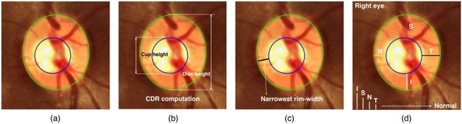
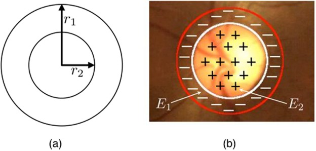
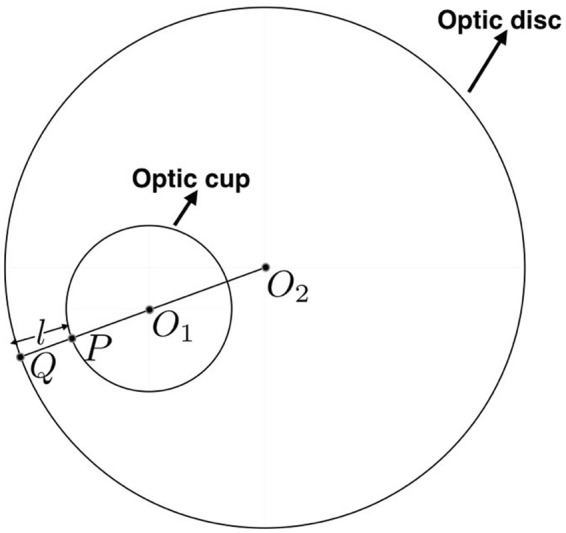
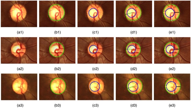
We present a novel and fully automated fundus image processing technique for glaucoma prescreening based on the rim-to-disc ratio (RDR). The technique accurately segments the optic disc and optic cup and then computes the RDR based on which it is possible to differentiate a normal fundus from a glaucomatous one. The technique performs a further categorization into normal, moderate, or severely glaucomatous classes following the disc-damage-likelihood scale (DDLS). To the best of our knowledge, this is the first engineering attempt at using RDR and DDLS to perform glaucoma severity assessment. The segmentation of the optic disc and cup is based on the active disc, whose parameters are optimized to maximize the local contrast. The optimization is performed efficiently by means of a multiscale representation, accelerated gradient-descent, and Green's theorem. Validations are performed on several publicly available databases as well as data provided by manufacturers of some commercially available fundus imaging devices. The segmentation and classification performance is assessed against expert clinician annotations in terms of sensitivity, specificity, accuracy, Jaccard, and Dice similarity indices. The results show that RDR based automated glaucoma assessment is about 8% to 10% more accurate than a cup-to-disc ratio (CDR) based system. An ablation study carried out considering the ground-truth expert outlines alone for classification showed that RDR is superior to CDR by 5.28% in a two-stage classification and about 3.21% in a three-stage severity grading.
Conflict of interest statement
The authors declare no competing interests.
Figures


References
-
- Giaconi, J. A., Law, S. K., Coleman, A. L. & Caprioli, J. Pearls of Glaucoma Management. Springer, USA (2010).
Publication types
MeSH terms
LinkOut - more resources
Full Text Sources
Medical
