. 2019 May;569(7757):503-508.
doi: 10.1038/s41586-019-1186-3. Epub 2019 May 8.

Next-generation characterization of the Cancer Cell Line Encyclopedia

Mahmoud Ghandi #  1 Franklin W Huang #  1   2   3 Judit Jané-Valbuena  1   2 Gregory V Kryukov  1 Christopher C Lo  1 E Robert McDonald 3rd  4 Jordi Barretina  4 Ellen T Gelfand  1 Craig M Bielski  1 Haoxin Li  1   2 Kevin Hu  1 Alexander Y Andreev-Drakhlin  1 Jaegil Kim  1 Julian M Hess  1 Brian J Haas  1 François Aguet  1 Barbara A Weir  1 Michael V Rothberg  1 Brenton R Paolella  1 Michael S Lawrence  1   5   6   7 Rehan Akbani  8 Yiling Lu  8 Hong L Tiv  9 Prafulla C Gokhale  9 Antoine de Weck  10 Ali Amin Mansour  1 Coyin Oh  1 Juliann Shih  1 Kevin Hadi  11   12 Yanay Rosen  1 Jonathan Bistline  1 Kavitha Venkatesan  4 Anupama Reddy  4 Dmitriy Sonkin  4   13 Manway Liu  4 Joseph Lehar  4 Joshua M Korn  4 Dale A Porter  4 Michael D Jones  4 Javad Golji  4 Giordano Caponigro  4 Jordan E Taylor  1 Caitlin M Dunning  1 Amanda L Creech  1 Allison C Warren  1 James M McFarland  1 Mahdi Zamanighomi  1 Audrey Kauffmann  10 Nicolas Stransky  1 Marcin Imielinski  11   12 Yosef E Maruvka  1   5 Andrew D Cherniack  1   2 Aviad Tsherniak  1 Francisca Vazquez  1 Jacob D Jaffe  1 Andrew A Lane  2 David M Weinstock  2 Cory M Johannessen  1 Michael P Morrissey  4 Frank Stegmeier  4 Robert Schlegel  4 William C Hahn  1   2 Gad Getz  1   5   6   7 Gordon B Mills  8 Jesse S Boehm  1 Todd R Golub  1   2   14 Levi A Garraway  1   2 William R Sellers  15   16
Affiliations

Next-generation characterization of the Cancer Cell Line Encyclopedia

Mahmoud Ghandi et al. Nature. 2019 May.

Abstract

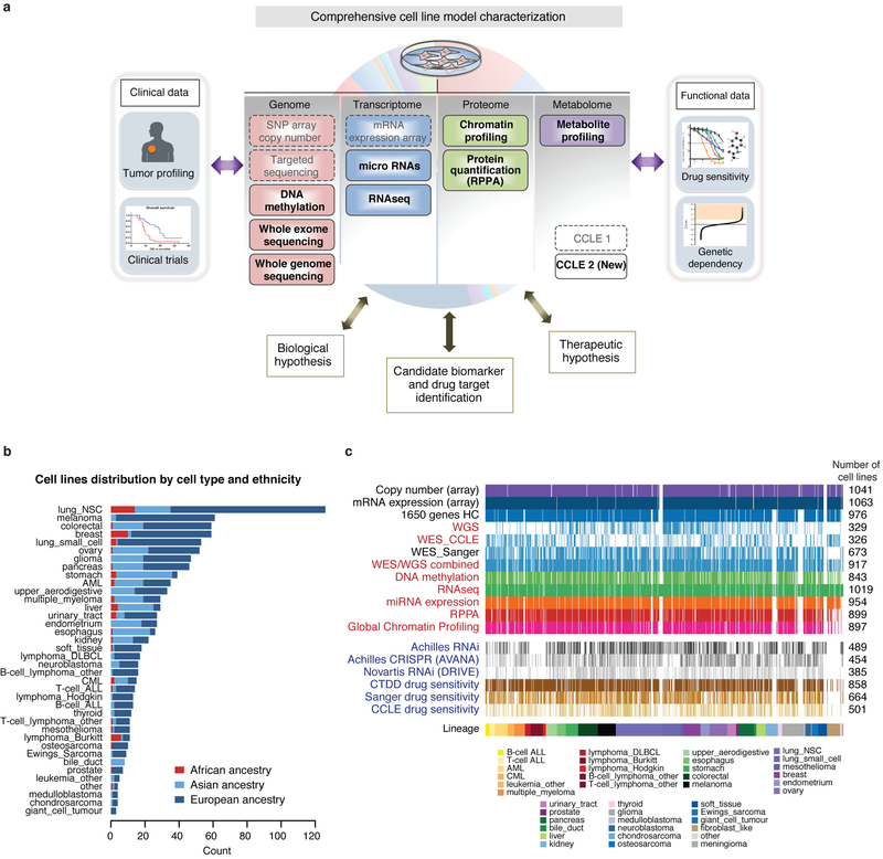
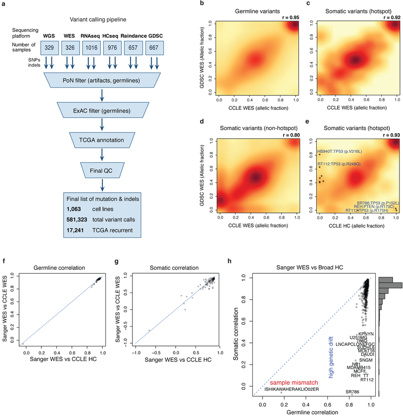
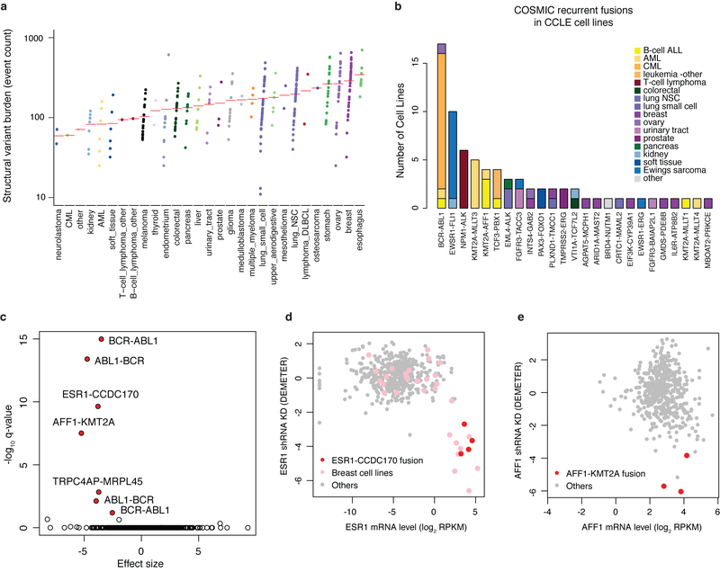
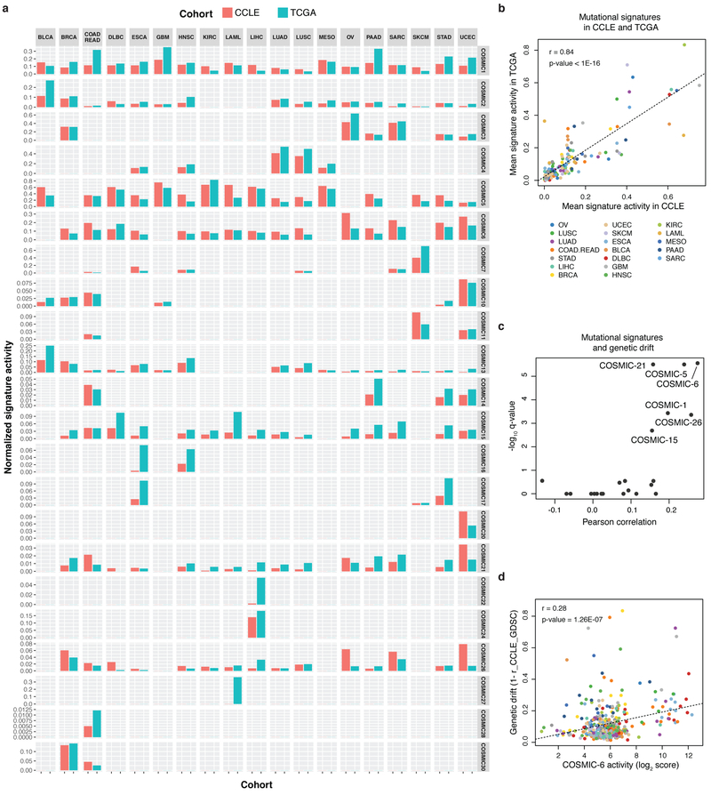
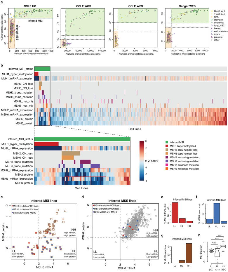
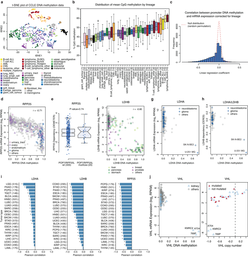
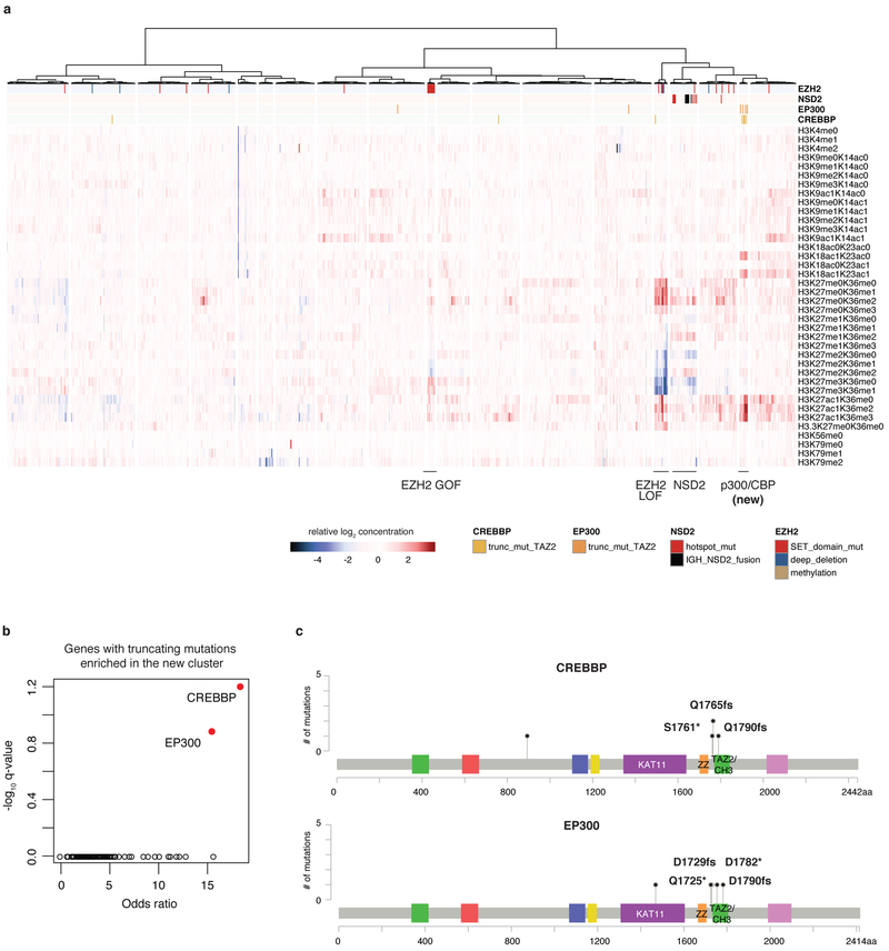
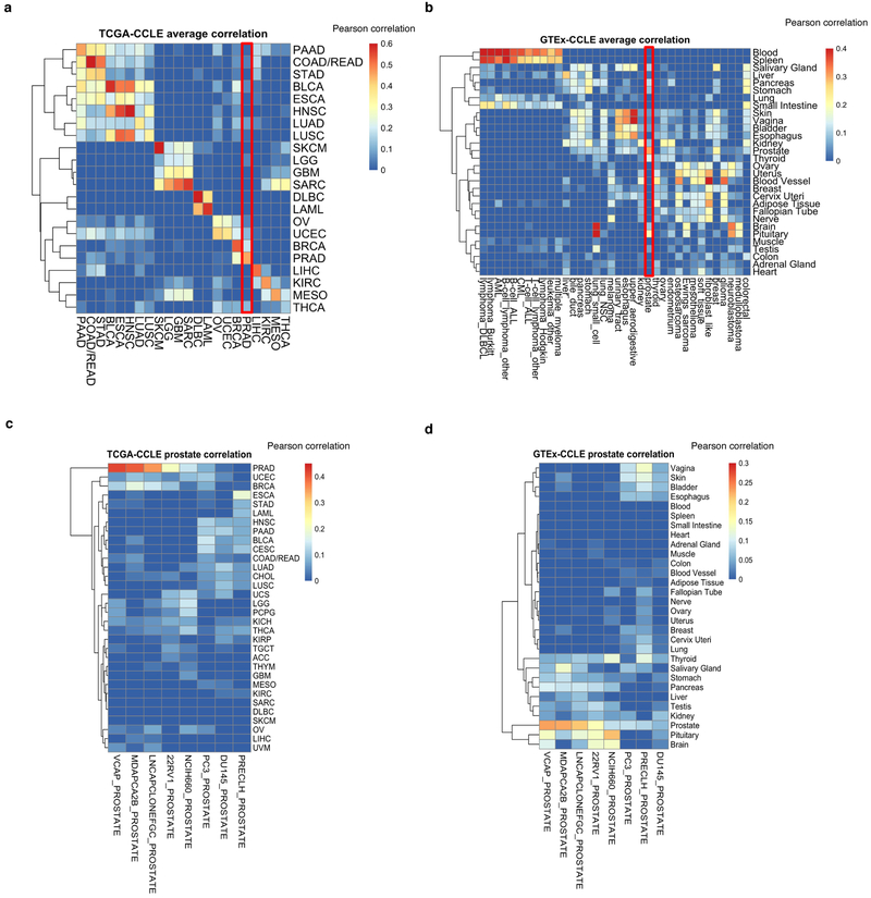
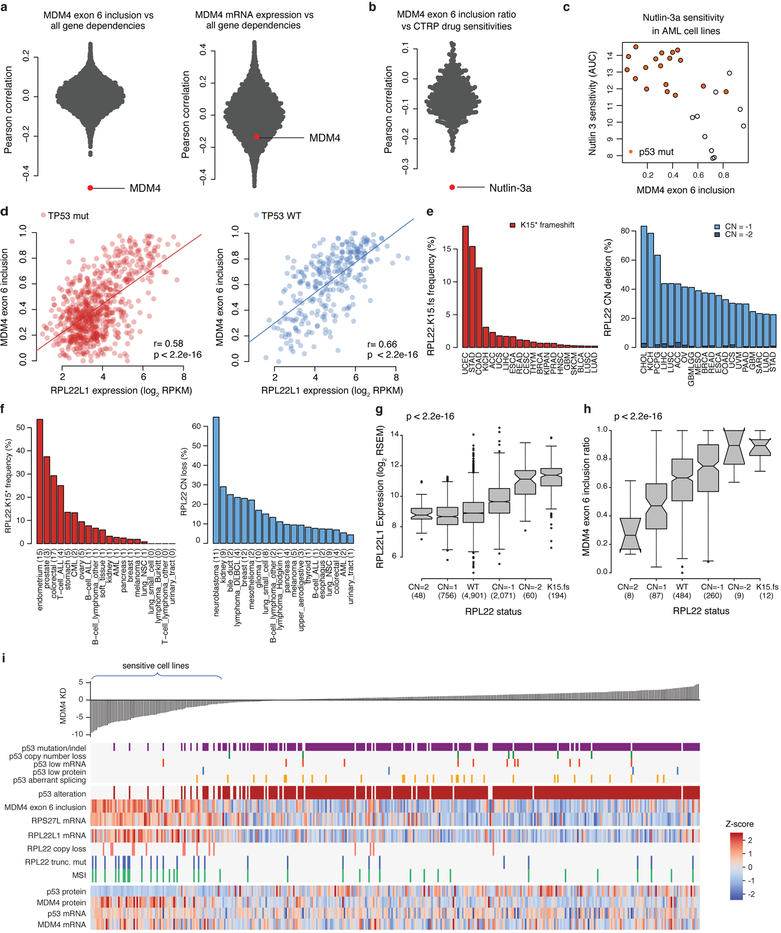
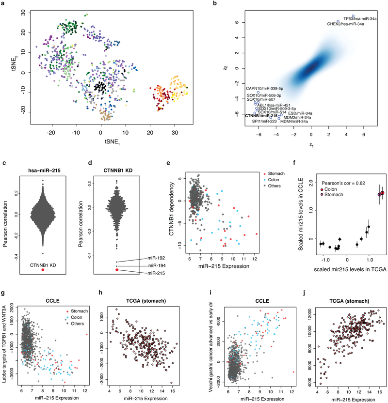
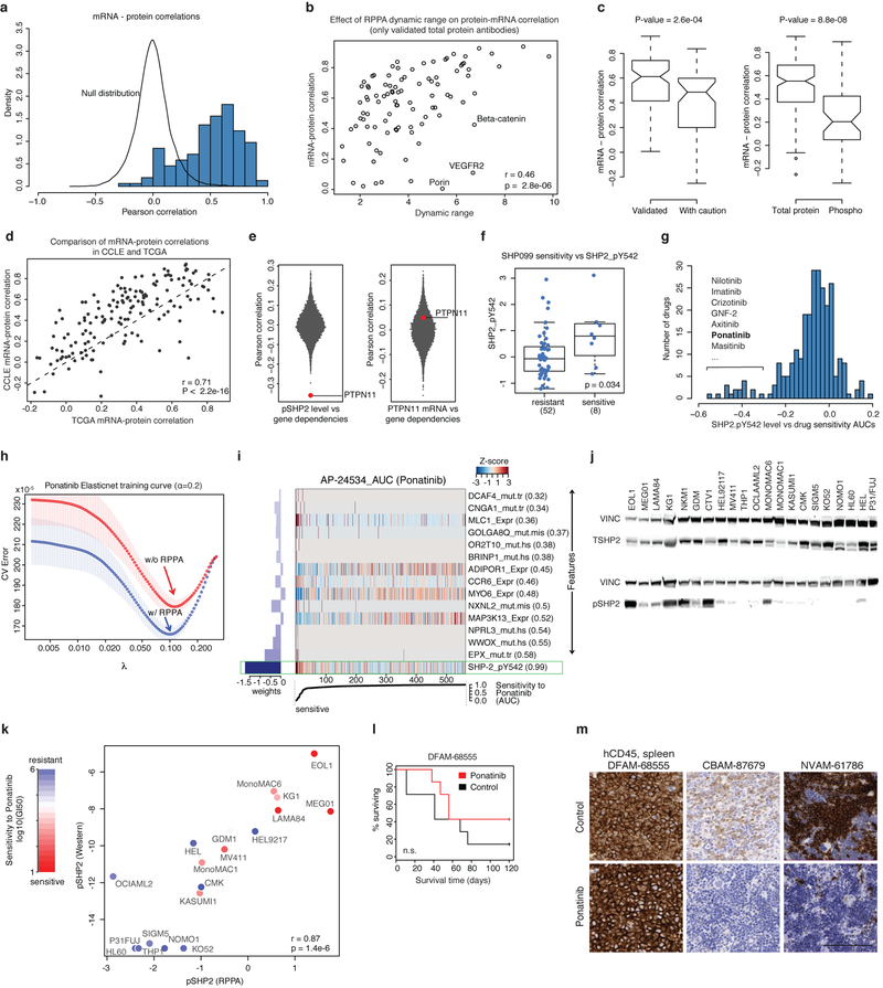
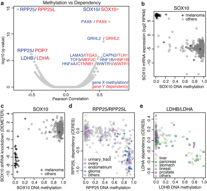
Large panels of comprehensively characterized human cancer models, including the Cancer Cell Line Encyclopedia (CCLE), have provided a rigorous framework with which to study genetic variants, candidate targets, and small-molecule and biological therapeutics and to identify new marker-driven cancer dependencies. To improve our understanding of the molecular features that contribute to cancer phenotypes, including drug responses, here we have expanded the characterizations of cancer cell lines to include genetic, RNA splicing, DNA methylation, histone H3 modification, microRNA expression and reverse-phase protein array data for 1,072 cell lines from individuals of various lineages and ethnicities. Integration of these data with functional characterizations such as drug-sensitivity, short hairpin RNA knockdown and CRISPR-Cas9 knockout data reveals potential targets for cancer drugs and associated biomarkers. Together, this dataset and an accompanying public data portal provide a resource for the acceleration of cancer research using model cancer cell lines.


Figures

Extended Data Fig. 1
Extended Data Fig. 1. Overview of CCLE cell lines and datasets.
a, The existing and new CCLE datasets as indicated are depicted. b, Distribution of cell lines by lineage and ancestry across CCLE. c, Visual representation of the number of cell lines in each dataset. New CCLE datasets are shown in red. Functional genomics datasets are shown in blue.
Extended Data Fig. 2
Extended Data Fig. 2. CCLE variant calling pipeline and CCLE and GDSC comparison.
a, Unified pipeline integrating mutation and indel calls from different platforms was used to generate a set of high confidence genomic alterations across 1,063 cancer cell lines. Identified variants were cross-referenced with the ExAC and TCGA databases and a panel of normals (PoN) to exclude germline variants/artefacts and generate the finalized high-confidence variant call set. bd, Comparison of variant calls between CCLE and Sanger GDSC cell lines for germline (b; n = 1,250,562), TCGA hotspot somatic (c; n = 281) and non-hotspot somatic (d; n = 82,572) variants using WES data. Pearson’s correlation coefficients are shown. e, Comparison of TCGA hotspot variant calls between CCLE Hybrid Capture (HC) data and Sanger GDSC WES data. Variants with allelic fraction >0.4 in one dataset and greater than fourfold difference in allelic fractions between the two datasets are shown as open circles (n = 980). f, g, Comparison of Pearson’s correlation coefficients between CCLE WES and Sanger GDSC WES data versus Pearson’s correlation coefficients between CCLE HC and Sanger GDSC WES data for germline (f; n = 107) and somatic (g; n = 93) variants. Cell lines with fewer than 30 variants were excluded. h, Comparison of allelic fraction Pearson’s correlations between CCLE cell lines and Sanger cell lines using CCLE HC and Sanger GDSC WES data (n = 558 common cell lines between the two datasets; Supplementary Table 3). Cell lines with low germline correlation (sample mismatch) and low somatic correlation (genetic drift) are highlighted.
Extended Data Fig. 3
Extended Data Fig. 3. Annotation of structural variants and fusions in CCLE cell lines.
a, Structural variant (SV) burden in CCLE whole genomes. Structural variants detected by SvABA in cell lines grouped by tissue type are plotted in the order of mean structural variant burden (red bar in each facet). b, Bar plot of recurrent COSMIC fusions detected in CCLE RNA-seq data coloured by cell line lineage. c, Volcano plot of Achilles RNAi gene dependencies versus CCLE fusions for cell lines (n = 478) common between CCLE and Achilles datasets. P values determined by two-sided t-test. Genes with significant adjusted P values (false discovery rate (FDR) < 0.1) are highlighted. d, e, Examples of fusions associated with gene dependency: cell lines with ESR1-CCDC170 fusion (n = 4) are sensitive to ESR1 shRNA knockdown (d), and cell lines with AFF1-KMT2A fusion (n = 3) are sensitive to AFF1 shRNA knockdown (e). The x axis shows mRNA expression, and the y axis shows Achilles RNAi gene dependency DEMETER score.
Extended Data Fig. 4
Extended Data Fig. 4. Comparison of COSMIC mutational signatures in CCLE and TCGA datasets.
a, Mutational signature activity for CCLE cell lines and TCGA tumours averaged for each cancer type. For each sample, we computed a fraction of mutations attributed to 30 COSMIC signatures and took average across samples in each cancer type. Tumour types selected for representation have at least 20 samples in CCLE. b, Scatterplots for the mutational signature activities for CCLE and TCGA (n = 168). P value determined by linear regression analysis and corrected for COSMIC signature number. c, Volcano plot for comparison of COSMIC mutational signatures and CCLE or GDSC genetic drift estimates using two-sided Pearson’s correlation test (n = 3–459; Supplementary Table 6). d, Scatter plot for COSMIC-6 mutational activity signatures versus CCLE or GDSC genetic drift estimates (n = 354). Colour coding as in b. P value determined by Pearson’s correlation test.
Extended Data Fig. 5
Extended Data Fig. 5. Determination of MSI status in the CCLE and interrogation of mismatch repair genes.
a, Identification of MSI cell lines. Number of deletions in microsatellite regions is plotted versus percentage of deletions in microsatellite regions for all cell lines in CCLE HC sequencing, CCLE WGS, CCLE WES, and Sanger GDSC WES datasets (see Methods). The x axis denotes the number of short deletions in microsatellite regions, and the y axis denotes percentage microsatellite as measured by the percentage of short deletions that lie within microsatellite regions. Inferred MSI cell lines are outlined by the green rectangle. b, Heat plot of inferred MSI status and selected CCLE annotations for DNA mismatch repair genes MLH1, MSH2 and MSH6 genes for all cell lines (top) and the MSI subset (bottom). Highlighted red boxes show differences in mRNA and protein expression levels in MSH2 and MSH6. MLH1 hypermethylation is defined as average promoter methylation greater than 0.5. c, d, Scatterplot of CCLE cell lines comparing MSH6 mRNA expression levels (x axis) from RNA-seq versus MSH6 protein abundance (y axis) as quantified by RPPA in inferred-MSI (c) and inferred-MSS (d) cell lines. Red and blue denotes cell lines containing truncating mutations or copy number loss in MSH6 and MSH2, respectively. Purple denotes cell lines containing truncating mutation or copy number loss in both MSH2 and MSH6. The black box highlights the MSH6 high mRNA low protein (HL) category. eg, Bar plots of percentages of cell lines containing truncating mutations in MSH6 (e), MSH2 (f), and MLH1 expression loss (g) in different MSH6 mRNA and protein categories among inferred-MSI cell lines (LL: n = 11; HL: n = 17; HH: n = 44). P = 4 × 10⁻⁴ (e), P = 1 × 10⁻³ (f) and P = 1 × 10⁻⁴ (g), two-sided Fisher test. h, MSH2 protein levels in different MSH6 mRNA and protein categories. ***P < 1 × 10⁻⁶, two-sided Wilcoxon rank sum test. P = 8 × 10⁻¹⁴, difference between the HH and HL set; P = 1 × 10⁻⁸, difference between the HH and LL set. Box plots as defined in Fig. 4d.
Extended Data Fig. 6
Extended Data Fig. 6. Examples of DNA methylation associated with gene expression and dependencies in cell lines.
a, t-SNE plot for DNA methylation data across all CCLE cell lines. Each dot represents a cell line coloured by cell lineage. b, Distribution of mean CpG methylation in CCLE cell lines (n = 843) grouped by cancer type. Box plots as defined in Fig. 4d. c, Correlation of promoter methylation and gene expression for all genes corrected for cancer type (n = 836 cell lines, 18,296 genes). The y axis represents the number of genes, and the x axis is the linear regression coefficient corresponding to normalized promoter DNA methylation. Cancer types were used as covariates in the linear regression analysis. A subset of genes show significant correlation between higher promoter methylation and lower gene expression (n = 7,388; permutation test P < 0.05; Methods). Dotted line shows the empirical null distribution. d, Cell lines with higher levels of RPP25 methylation show decreased RPP25 mRNA expression (Pearson’s r = −0.79, n = 834 cell lines; P < 2.2 × 10⁻¹⁶). e, Comparison of Achilles RNAi RPP25 gene dependency scores for cell lines with and without truncating mutation or copy number loss in POP7 or RPP25L genes (n = 458 cell lines; P = 0.74, two-sided Wilcoxon rank sum test). Box plots as defined in Fig. 4d. f, Cell lines with higher levels of LDHB methylation show decreased LDHB mRNA expression (Pearson’s r = −0.80, n = 815 cell lines; P < 2.2 × 10⁻¹⁶). g, Cell lines with higher levels of LDHA methylation show decreased LDHA expression. Two cell lines, SK-N-BE2 and U-251-MG, show markedly higher LDHA methylation and decreased LDHA expression (Pearson’s r = −0.27, n = 836; P = 5.34 × 10⁻¹⁶). h, Cell lines with high levels of LDHA methylation display sensitivity to LDHB knockout by CRISPR–Cas9 screening (Pearson’s r = −0.53, n = 371, P < 2.2 × 10⁻¹⁶). i, Promoter methylation versus mRNA expression correlations in TCGA tumour types (sample sizes shown in parentheses). *P < 0.001, Pearson’s correlation test. j, Scatterplot of CCLE lines comparing expression of tumour suppressor VHL (Von Hippel-Landau) mRNA versus VHL methylation (left, all cell lines) and copy number (right, kidney subset). VHL hypermethylation in three kidney cell lines is associated with marked loss of VHL expression. VHL is inactivated by DNA copy number loss, somatic mutation, and promoter hypermethylation.
Extended Data Fig. 7
Extended Data Fig. 7. Global chromatin profiling dataset.
a, Unsupervised clustering of global chromatin profiling data for 897 CCLE cell lines. Each column corresponds to an individual cell line and each row corresponds to a specific combination of chromatin post-translational modifications (‘marks’). For each mark, the fold change relative to median of cell lines is depicted on the heat map. EZH2, NSD2, CREBBP and EP300 status are annotated. Previously described clusters (associated with EZH2 gain of function, EZH2 loss of function, and NSD2 alterations), as well as the newly identified cluster associated with p300 and CBP gain-of-function alterations, are annotated. b, Volcano plot for truncating mutation enrichment analysis in the newly identified cluster, characterized by marked increases in H3K18 and H3K27 acetylation is shown (n = 893 cell lines; adjusted P values determined by two-sided Fisher’s exact test). EP300 and CREBBP are the top two genes with truncating mutations enriched in this cluster. Only genes with at least 20 affected cell lines (n = 684 genes) were included. c, Distribution of truncating mutations affecting EP300 and CREBBP in the 10 cell lines in the newly identified p300/CBP cluster. Truncating mutations predicted to affect the TAZ2 (CH3) domain specifically are highlighted. Two other truncating mutations not specific to TAZ2 (CH3) are OVCAR-8 (S893*) and COLO-704 (K1469fs).
Extended Data Fig. 8
Extended Data Fig. 8. Comparison of CCLE gene expression data with primary tumour (TCGA) and normal tissue (GTEX) gene expression datasets.
a, Comparison of gene expression profiles between the CCLE cell lines (n = 1,019) and TCGA primary tumours (n = 10,535). For every gene in each dataset, expression values were averaged per cancer type and then mean centred across types. Pearson correlation values were calculated between the CCLE and TCGA cancer types using the (n = 5,000) most highly variable genes. b, Comparison of average gene expression profiles between the CCLE cell lines (n = 1,019) and the GTEx normal tissues (n = 11,688). Similar to a, expression profiles for each tissue type in GTEx was correlated with the CCLE expression profiles (n = 5,000 genes). c, Gene expression comparison of eight prostate cell lines and TCGA primary tumour samples (n = 5,000 genes). d, Gene expression comparison of eight prostate cell lines and GTEx normal tissue samples (n = 5,000 genes).
Extended Data Fig. 9
Extended Data Fig. 9. MDM4 alternative splicing and association with RPL22 and RPL22L1.
a, Distribution of MDM4 exon 6 inclusion (left) and MDM4 mRNA expression (right) correlation with all gene dependencies in the Achilles RNAi dataset (n = 189–478; Supplementary Table 10). b, Correlation of MDM4 exon 6 inclusion with sensitivity to all small molecules in the CTRP AUC dataset using all cell lines. Nutlin-3a is the top drug sensitivity correlated with MDM4 exon 6 inclusion (n = 79–810; Supplementary Table 10). c, Example of nutlin-3a sensitivity versus MDM4 exon 6 inclusion in the AML cell lines (Spearman correlation rho = −0.64, P = 3 × 10⁻⁴, n = 28). The y axis shows AUC for nutlin-3a in the CTRP dataset. d, Scatterplot of MDM4 exon 6 inclusion versus RPL22L1 expression for all p53-mutant (left, n = 711) and p53 wild-type (right, n = 288) CCLE cell lines. P values determined by Pearson’s correlation test. e, Frequency of RPL22 recurrent frameshift mutations (left) and copy number deletions (right) in TCGA. f, Frequency of RPL22 recurrent frameshift mutations (left) and copy number deletions (right) in CCLE. g, Correlation of RPL22L1 mRNA expression with RPL22 copy number loss and RPL22 frameshift deletions in TCGA. P value determined by two-sided Kruskal–Wallis rank sum test. Box plots as defined in Fig. 4d. Values in parentheses denote sample size in each category. h, Correlation of MDM4 exon 6 inclusion with RPL22 copy number loss and RPL22 frameshift deletions in TCGA. P value determined by two-sided Kruskal–Wallis rank sum test. Box plots are as defined in Fig. 4d. Values in parentheses denote sample size in each category. i, Selected genomic features that correlate with sensitivity to MDM4 shRNA knockdown. mRNA expression of MDM4 and TP53 are shown for comparison.
Extended Data Fig. 10
Extended Data Fig. 10. Examples of microRNA expression associated with gene dependencies in cell lines.
a, t-SNE plot for miRNA data across all CCLE cell lines. Each dot represents a cell line. Each colour represents a different cell lineage. Colour coding is as in Fig. 1. b, Scatter plot of pairwise Pearson’s correlation of gene dependency and miRNA expression (n = 420 cell lines), normalized for each microRNA (z1, x axis) and each gene dependency (z2, y axis). Strong outlier pairs with |z1|>6 or |z2|>6 are highlighted. c, Distribution of Pearson’s correlations of mir-215 expression with Achilles RNAi gene dependencies for 16,871 genes (n = 162–420 cell lines; Supplementary Table 13). CTNNB1 knockdown is the top negative correlate with mir-215 expression. d, Distribution of Pearson’s correlations of CTNNB1 gene dependency with all 734 measured miRNAs (n = 420 cell lines). The expression of mir-215 is the top gene negatively correlated with CTNNB1 dependency. mir-215 and mir-194–1 cluster together at 1q41, whereas mir-192 and mir-194–2 cluster at 11q13.1. mir-215 and mir-192 are close homologues. e, Scatterplot of mir-215 expression versus CTNNB1 dependency of all CCLE cell lines. Colon and stomach lineages are shown in blue and red, respectively. f, Scaled mir-215 expression in TCGA and CCLE datasets (n = 14; mean ± s.e.m.). Stomach and colorectal lineages in both datasets have high mir-215 expression. g, Single-sample gene set enrichment analysis identifies TGFB1 and WNT3A pathway gene sets correlated with mir-215 expression using CCLE RNA-seq data. The gene set ‘Labbe targets of TGFB1 and WNT3A’ of downstream targets of TGF-β and WNT ligands is negatively correlated with mir-215 expression. h, The gene set ‘Labbe targets of TGFB1 and WNT3A’ is negatively correlated with mir-215 expression in the TCGA stomach mRNA expression dataset. i, The gene set ‘Vecchi gastric advanced vs early dn’ of down-regulated genes distinguishing between advanced and early gastric cancer subtypes is positively correlated with mir-215 expression in the CCLE. j, mir-215 expression in the stomach TCGA mRNA expression dataset is positively correlated with the ‘Vecchi gastric advanced vs early dn’ gene set.
Extended Data Fig. 11
Extended Data Fig. 11. RPPA analysis.
a, Distribution of Pearson’s correlation coefficient between total protein levels as measured by RPPA and mRNA expression levels measured by RNA-seq (n = 890 cell lines, 154 genes). The empirical null distribution for correlation of mRNA and protein for two random genes is shown for comparison (P < 2.2 × 10⁻¹⁶, two-sided Wilcoxon rank sum test). b, Effect of RPPA dynamic range on mRNA and protein correlation (n = 96). mRNA and protein correlation is plotted against dynamic range for each validated total protein antibody. Most antibodies with low mRNA and protein correlation tend to have low dynamic range with the exception of VEGFR2 gene, which despite high dynamic range, exhibits very low mRNA and protein correlation. P values determined by two-sided Pearson’s correlation test. c, Effect of RPPA antibody quality and target type on mRNA/protein correlation. On the left, mRNA/protein Pearson correlation is plotted for ‘validated’ (n = 96) and ‘with caution’ (n = 58) antibodies for antibodies against total proteins. On the right, mRNA and protein Pearson’s correlation is plotted for antibodies against total protein (n = 154) and antibodies against phospho-protein (n = 50). Median correlations are 0.62 (validated), 0.48 (caution), 0.54 (total protein), 0.21 (phospho-protein). P values determined by two-sided Wilcoxon rank sum test. Box plots are as defined in Fig. 4d. d, Comparison of mRNA and protein correlations in CCLE and TCGA (n = 152). The Pearson’s correlation between mRNA and protein levels is calculated for each RPPA antibody in CCLE and TCGA separately. Each dot represents an antibody. Generally, the antibodies with low mRNA and protein correlation in CCLE also have low mRNA and protein correlation in TCGA data. P values determined by two-sided Pearson’s correlation test. e, Distribution of gene dependency (Achilles RNAi) correlations with RPPA pSHP2 level (left, n = 161–411, Supplementary Table 14) and PTPN11 mRNA expression (right, n = 192–478, Supplementary Table 14). PTPN11 dependency is strongly correlated with pSHP2 level, whereas there is no significant correlation with PTPN11 mRNA level. f, Comparison of pSHP2 levels in SHP099-sensitive and -resistant cell lines (n = 60). P value determined by two-sided Wilcoxon rank sum test. SHP099 sensitivity data were obtained from ref.. Box plots are as defined in Fig. 4d. g, Pearson’s correlation of pSHP2 and Sanger GDSC drug sensitivity AUC dataset (n = 265 drugs and 198–588 overlapping cell lines). h, Model error for elastic net model of sensitivity to ponatinib with and without using RPPA data as predictive features. The y axis shows the cross-validation error (fivefold cross validation) against parameter β of elastic net (parameter β is fixed at 0.2). Data are mean ± s.d. for the five cross validation (CV) sets. The minimum CV error for models with and without using RPPA data are shown by arrows. i, Elastic net results for sensitivity to ponatinib. pSHP2 is the top feature selected by elastic net. On the left, elastic net weights (averaged over 200 bootstrapping trials) and colour-coded by the frequency each feature was selected by elastic net. The numbers in parentheses are the frequency each feature was selected. Each column is a cell line and each row is a feature. The cell lines are sorted by their sensitivity to ponatinib (shown at the bottom). j, Western blot analysis of pSHP2 and total SHP2 levels across AML and select CML cell lines. Western blots were performed twice independently with similar results. k, Validation of RPPA data for pSHP2. pSHP2 levels measured by western blot are plotted against pSHP2 levels measured by RPPA for the tested AML and control CML cell lines (n = 19). The cell lines are colour-coded by their sensitivity to ponatinib. P values determined by two-sided Pearson’s correlation test. l, In vivo mouse xenograft experiment survival curves of ponatinib-treated and control mice for the low pSHP2 primagraft DFAM-68555. (n = 7 mice in each treatment group). P values determined by log-rank (Mantle-Cox) test. m, Immunohistochemistry of spleen specimens from mice treated with control or ponatinib for 5 days using anti-CD45. Similar results were found using the other two independent sets of mice.
Fig. 1
Fig. 1. Overview of the datasets.
Representative heat maps from the CCLE datasets (n = 749). Cell lines grouped by cancer type; cancer types ordered by an unsupervised hierarchical clustering of mean values of each cancer type. From each dataset, a representative subset is shown, including mutation and fusion status in the top recurrently mutated genes and TERT promoter mutation, columns were randomly selected from CCLE copy number, DNA methylation, mRNA expression, exon inclusion, miRNA, protein array and global chromatin profiling datasets. Inferred-MSI status, inferred-ploidy and inferred-ancestries are shown. Unknown TERT promoter status is shown in light grey. AML, acute myeloid leukaemia; CML, chronic myelogenous leukaemia; ALL, acute lymphoid leukaemia; DLBCL, diffuse large B-cell lymphoma; NSC, non-small cell.
Fig. 2
Fig. 2. DNA methylation and cancer dependence.
a, Global correlation between DNA methylation and gene dependency of the same gene or associated genes (StringDB). Top pairs (q < 5 × 10⁻⁵) are labelled (n = 45–380; Supplementary Table 8). b, c, Hypomethylation of SOX10 in melanoma cell lines is associated with SOX10 mRNA expression (Pearson’s r = −0.82, n = 824, P < 2.2 × 10⁻¹⁶) (b) and sensitivity to SOX10 knockdown (Pearson’s r = 0.79, n = 376, P < 2.2 × 10⁻¹⁶) (c). RPKM, reads per kilobase of transcript per million mapped reads. d, Promoter hypermethylation of RPP25 is a marker for vulnerability to RPP25L knockout (Pearson’s r = −0.71, n = 369, P < 2.2 × 10⁻¹⁶). e, LDHB methylation confers sensitization to LDHA knockout (Pearson’s r = −0.52, n = 362, P < 2.2 × 10⁻¹⁶).
Fig. 3
Fig. 3. Global chromatin profiling reveals activating mutations in p300 and CBP.
A selected subset of the CCLE global chromatin profiling dataset showing H3K18 and H3K27 modifications in four clusters is shown from the unsupervised clustering of 897 cell lines. Each column represents a cell line, and each row a specific set of chromatin post-translational modifications (‘marks’). For each mark, the fold change relative to the median of cell lines is depicted. The new p300 and CBP cluster with acetylation marks are shown in bold. GOF, gain of function; LOF, loss of function.
Fig. 4
Fig. 4. MDM4 exon 6 inclusion is associated with MDM4 dependency and RPL22 or RPL22L1 status.
a, Scatterplot of correlation of gene dependency and exon inclusion (x axis) and correlation of gene dependency and gene expression (y axis) (n = 243,288 exons, 200–478 common cell lines; Supplementary Table 10; highlighted genes: |r_exon_inclusion| > 0.4). b, Alternative splicing generates two major MDM4 isoforms—full-length MDM4 (MDM4-FL) includes exon 6, whereas short MDM4 (MDM4-S) skips this exon. c, Validation of MDM4 exon 6 inclusion in a subset of CCLE cell lines (n = 16) using quantitative PCR (qPCR). Data are mean and s.d. of the log2(MDM4-FL/MDM4-S) ratio relative to the TOV21G standard cell line calculated across three technical replicates. d, e, Sensitivity of cell lines to MDM4 knockdown (DEMETER dependency scores) (d) and treatment with nutlin-3a (Cancer Therapeutics Response Portal (CTRP) area under the dose–response curve (AUC) scores) (e) by p53 mutational status (WT, wild type; mut, mutated) and the MDM4 splicing categories MDM4-S (MDM4 exon 6 inclusion ratio < 0.25) and MDM4-FL (inclusion ratio > 0.35). Numbers in parentheses denote the number of cell lines in each category. Box plots depict median (centre line), interquartile range (box), smaller of 1.5 times the interquartile range from the box, the minimum–maximum range (whiskers), and outliers (circles). f, Correlation of MDM4 exon 6 inclusion with gene expression (n = 1,003 cell lines). g, Correlation of RPL22L1 expression with exon-inclusion ratios (n = 200–1,019; Supplementary Table 10). P values determined by two-sided Spearman’s correlation test. h, i, Higher RPL22L1 expression (h) and MDM4 exon 6 inclusion (i) are associated with RPL22 copy number (CN) loss and RPL22 truncating mutations or indels. Box plots as defined in d. j, Scatterplot of RPL22L1 dependency versus RPL22L1 mRNA expression. Cell lines containing RPL22 truncating mutations and TP53 mutations are shown (n = 447). P values determined by two-sided Wilcoxon rank sum test (d, e, j), two-sided Spearman’s correlation test (f) or two-sided Kruskal–Wallis rank sum test (h, i).
Fig. 5
Fig. 5. High pSHP2 is a marker of SHP2 dependence and sensitivity to RTK inhibitors.
a, Global correlations of gene dependency and gene expression (y axis) versus correlation of gene dependency and protein expression. PTPN11 dependency is correlated with pSHP2 expression (Pearson’s r = −0.36, n = 411; P = 4.9 × 10⁻¹⁴) but not with mRNA expression (Pearson’s r = −0.07, n = 478; P = 0.15). b, A subset of AML lines (n = 21) show high pSHP2 expression associated with sensitivity to ponatinib. c, Validation of Sanger GDSC ponatinib sensitivity data in AML (n = 16) and CML (n = 2) cell lines. x axis is sensitivity to ponatinib in the Sanger GDSC dataset; y axis is sensitivity to ponatinib measured by CellTiter-Glo (CTG) cell viability assay. Each dot represents a cell line coloured by pSHP2 over total SHP2 level. IC50, half-maximal inhibitory concentration. d, In vitro validation of association of pSHP2 expression with sensitivity to ponatinib. Cell lines are annotated for known oncogenic events in the RTK pathway. tSHP2, total SHP2. e, pSHP2 levels measured by RPPA in mouse primagraft AML models (n = 14) and control cell lines (n = 6). Three models (bold) were chosen for in vivo validation experiments. f, In vivo mouse xenograft experiment survival curves. Ponatinib treatment prolonged survival in two primagrafts with high pSHP2 levels—CBAM-87679 and NVAM-61786—but not in the low pSHP2 primagraft DFAM-68555 (Extended Data Fig. 11l) (n = 7 mice in each group). P values determined by two-sided Pearson correlation test (ac) log-rank (Mantle–Cox) test (f).

References

    1. Barretina J et al. The Cancer Cell Line Encyclopedia enables predictive modelling of anticancer drug sensitivity. Nature 483, 603–607 (2012). - PMC - PubMed
    1. Li H et al. The Landscape of Cancer Cell Line Metabolism. Nat. Med (2019). [Accepted, 10.1038/s41591-019-0404-8] - DOI - PMC - PubMed
    1. Iorio F et al. A landscape of pharmacogenomic interactions in cancer. Cell 166, 740–754 (2016). - PMC - PubMed
    1. Ben-David U et al. Genetic and transcriptional evolution alters cancer cell line drug response. Nature 560, 325–330 (2018). - PMC - PubMed
    1. Tsherniak A et al. Defining a cancer dependency Map. Cell 170, 564–576.e516 (2017). - PMC - PubMed

Publication types