. 2019 Apr 24:10:401.
doi: 10.3389/fphys.2019.00401. eCollection 2019.

The Mechanical Stimulation of Myotubes Counteracts the Effects of Tumor-Derived Factors Through the Modulation of the Activin/Follistatin Ratio

Affiliations

The Mechanical Stimulation of Myotubes Counteracts the Effects of Tumor-Derived Factors Through the Modulation of the Activin/Follistatin Ratio

Alexandra Baccam et al. Front Physiol. .

Abstract

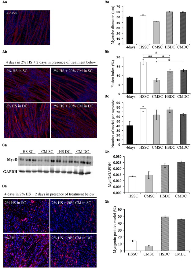
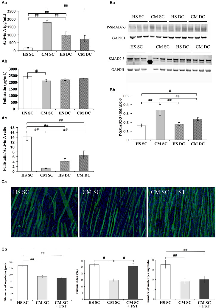
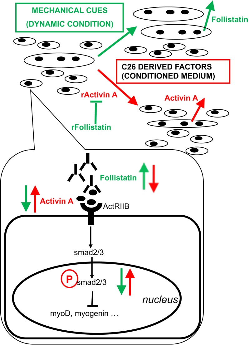
Activin negatively affects muscle fibers and progenitor cells in aging (sarcopenia) and in chronic diseases characterized by severe muscle wasting (cachexia). High circulating activin levels predict poor survival in cancer patients. However, the relative impact of activin in mediating muscle atrophy and hampered homeostasis is still unknown. To directly assess the involvement of activin, and its physiological inhibitor follistatin, in cancer-induced muscle atrophy, we cultured C2C12 myotubes in the absence or in the presence of a mechanical stretching stimulus and in the absence or presence of C26 tumor-derived factors (CM), so as to mimic the mechanical stimulation of exercise and cancer cachexia, respectively. We found that CM induces activin release by myotubes, further exacerbating the negative effects of tumor-derived factors. In addition, mechanical stimulation is sufficient to counteract the adverse tumor-induced effects on muscle cells, in association with an increased follistatin/activin ratio in the cell culture medium, indicating that myotubes actively release follistatin upon stretching. Recombinant follistatin counteracts tumor effects on myotubes exclusively by rescuing fusion index, suggesting that it is only partially responsible for the stretch-mediated rescue. Therefore, besides activin, other tumor-derived factors may play a significant role in mediating muscle atrophy. In addition to increasing follistatin secretion mechanical stimulation induces additional beneficial responses in myotubes. We propose that in animal models of cancer cachexia and in cancer patients purely mechanical stimuli play an important role in mediating the rescue of the muscle homeostasis reported upon exercise.

Keywords: C26 colon carcinoma; FlexCell apparatus; exercise; mechanotransduction; myokines; skeletal muscle atrophy.


Figures

FIGURE 1
FIGURE 1
Mechanical stimulation counteracts the negative effect of tumor-derived factors. (A) Myosin (red) localization and nuclei (blue) by immunofluorescence in C2C12 myotubes at 4d (Aa) and 6d (Ab) of culture in a differentiation medium in the absence (HS) or presence (CM) of C26-conditioned medium, in combination with the absence (SC) or presence (DC) of cyclic stretching. (B) Morphometric analyses were performed on replicate samples (n = 6). One-way ANOVA performed on data from 4d and 6d (five groups) followed by Dunnet’s test indicated a significant increase in the fusion index (FI) and in the number of nuclei/myotube (NpM) between 4d and 6d in any condition except CM. Two-way ANOVA performed on 6d values showed a significant effect for: (Ba) DC on myotube diameter (F = 12.66; df = 1; p < 0.05); (Bb) CM and interaction with DC (for CM: F = 24.73; df = 1; p < 0.001; for interaction: F = 30.2; df = 1; p < 0.001) on fusion index; #p < 0.05, ##p < 0.01 by Tukey HSD test); (Bc) CM on number of nuclei/myotube (F = 5.64; df = 1; p < 0.05). (C) WB analysis for MyoD (Ca) and relative average density (Cb) following normalization over the GAPDH signal in the same conditions as above. Two-way ANOVA showed a significant effect for DC (F = 19.47; df = 1; p < 0.001). (D) Myogenin (red) and nuclei (blue) by IF (Da) in C2C12 myotubes at 6d of culture in the same conditions as above, and quantification of the percentage of myogenin+ nuclei (Db). Two-way ANOVA showed the significant effect of both CM (F = 13.55; df = 1; p < 0.01) and DC (F = 451.7; df = 1; p < 0.0001), indicating that the DC rescues myogenin expression. Data are shown as mean ± SEM.
FIGURE 2
FIGURE 2
Follistatin is not sufficient to rescue myotube size in the presence of tumor-derived factors. (A) Quantification of activin (Aa) and follistatin (Ab) by ELISA in 6d culture supernatant, in differentiation medium in the absence (HS) or presence (CM) of C26-conditioned medium, in combination with the absence (SC) or presence (DC) of cyclic stretch. The follistatin/activin ratio was calculated (Ac). Two-way ANOVA showed: CM effect and a interaction with DC on activin levels; interaction between CM and DC on follistatin levels; CM effect and interaction with DC on follistatin/activin ratio. #p < 0.05, ##p < 0.01 by Tukey HSD test. (B) WB analysis for P-SMAD2/3 and SMAD 2/3 (Ba) and relative average density (Bb) following normalization over the GAPDH. Two-way ANOVA showed a significant effect for DC (F = 6.25; df = 1; p < 0.05) and interaction of Dc with CM (F = 12.36; df = 1; p < 0.01). #p < 0.05, ##p < 0.01 by Tukey HSD test. (C) Immunofluorescence for Myosin (green) and nuclei (blue) in C2C12 myotubes at 6d of culture (Ca) in differentiation medium in the absence (HS) or presence (CM) of C26-conditioned medium and in CM supplemented with 100 ng/ml recombinant follistatin, in the absence of cyclic stretch (SC). The treatments were performed starting on 4d culture and changing the medium daily. Morphometric analyses (Cb) were performed on replicate samples (n = 6). One-way ANOVA indicated a significant effect of treatments on myotube diameter and on the number of nuclei/myotube. ∗∗p < 0.01 by Tukey HSD test.
FIGURE 3
FIGURE 3
Proposed model of action of tumor-derived factors and mechanical stimulation on myotubes and myoblasts. Mixed cultures of myotubes and myoblasts mature in culture by increasing the diameter of myotubes, the fusion index (i.e., myogenic differentiation tout court, including the formation of novel myotubes) and the number of nuclei per myotube (i.e., myotube accretion by incorporation of myoblasts). C26 tumor-derived factors include activin and induce further expression and release of activin as well as a decrease of follistatin expression and its release by muscle cells, ultimately leading to myotube atrophy, a block of myogenic differentiation and hampered incorporation of myoblasts into myotubes. On the other hand, mechanical stimulation counteracts the negative effects exerted by tumor-derived factors on muscle cells by diminishing the levels of activin available to bind actRIIB: this is obtained by reducing activin concentration in the medium and by rescuing follistatin release by muscle cells. Recombinant activin (rActivin A) mimics tumor CM and its effects are counteracted by recombinant follistatin (rFollistatin). However, rFollistatin only partially counteracts CM: since, in the presence of CM, follistatin rescues the fusion index but not myotube diameter nor the number of NpM, while mechanical stimulation also reverts CM-mediated effects on myotube size, follistatin is mostly responsible for the regulation of myogenic differentiation, while mechanical stimulation preserves myotube size through additional mechanisms. The signaling pathways downstream of actRIIB involve the activation of SMAD2/3 transcriptional activity, which is increased by tumor-derived factors and decreased by mechanical stimulation, resulting in the regulation of MRF expression leading to myoblast differentiation and fusion.

References

    1. Aulino P., Costa A., Chiaravalloti E., Perniconi B., Adamo S., Coletti D., et al. (2015). Muscle extracellular matrix scaffold is a multipotent environment. Int. J. Med. Sci. 12 336–340. 10.7150/ijms.10761 - DOI - PMC - PubMed
    1. Barone R., Macaluso F., Sangiorgi C., Campanella C., Marino Gammazza A., Moresi V., et al. (2016). Skeletal muscle Heat shock protein 60 increases after endurance training and induces peroxisome proliferator-activated receptor gamma coactivator 1 alpha1 expression. Sci. Rep. 6:19781. 10.1038/srep19781 - DOI - PMC - PubMed
    1. Barreto R., Mandili G., Witzmann F. A., Novelli F., Zimmers T. A., Bonetto A. (2016). Cancer and chemotherapy contribute to muscle loss by activating common signaling Pathways. Front. Physiol. 7:472. - PMC - PubMed
    1. Carotenuto F., Coletti D., Di Nardo P., Teodori L. (2016). alpha-linolenic acid reduces TNF-induced apoptosis in C2C12 myoblasts by regulating expression of apoptotic proteins. Eur. J. Transl. Myol. 26:6033. 10.4081/ejtm.2016.6033 - DOI - PMC - PubMed
    1. Chen J. L., Colgan T. D., Walton K. L., Gregorevic P., Harrison C. A. (2016). The TGF-beta signalling network in muscle development, adaptation and disease. Adv. Exp. Med. Biol. 900 97–131. 10.1007/978-3-319-27511-6_5 - DOI - PubMed

LinkOut - more resources