. 2019 Apr 24:10:413.
doi: 10.3389/fphys.2019.00413. eCollection 2019.

Glucagon Receptor Signaling and Lipid Metabolism

Affiliations

Glucagon Receptor Signaling and Lipid Metabolism

Katrine D Galsgaard et al. Front Physiol. .

Abstract

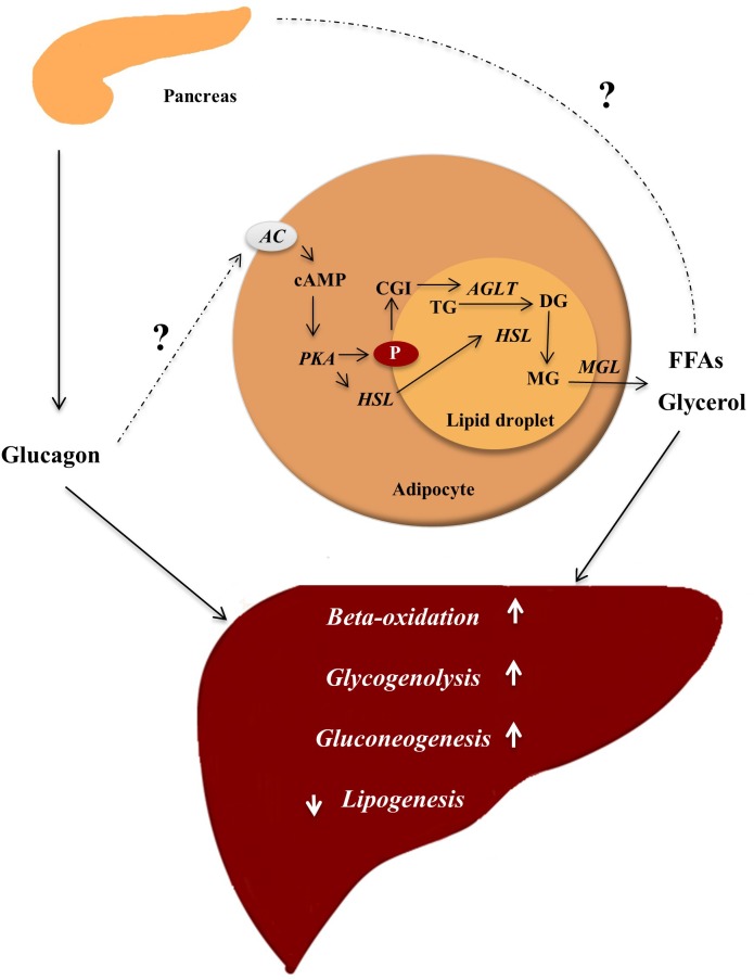
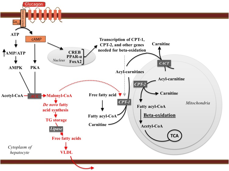
Glucagon is secreted from the pancreatic alpha cells upon hypoglycemia and stimulates hepatic glucose production. Type 2 diabetes is associated with dysregulated glucagon secretion, and increased glucagon concentrations contribute to the diabetic hyperglycemia. Antagonists of the glucagon receptor have been considered as glucose-lowering therapy in type 2 diabetes patients, but their clinical applicability has been questioned because of reports of therapy-induced increments in liver fat content and increased plasma concentrations of low-density lipoprotein. Conversely, in animal models, increased glucagon receptor signaling has been linked to improved lipid metabolism. Glucagon acts primarily on the liver and by regulating hepatic lipid metabolism glucagon may reduce hepatic lipid accumulation and decrease hepatic lipid secretion. Regarding whole-body lipid metabolism, it is controversial to what extent glucagon influences lipolysis in adipose tissue, particularly in humans. Glucagon receptor agonists combined with glucagon-like peptide 1 receptor agonists (dual agonists) improve dyslipidemia and reduce hepatic steatosis. Collectively, emerging data support an essential role of glucagon for lipid metabolism.

Keywords: adipose tissue; alpha cell; glucagon; lipid; liver.


Figures

FIGURE 1
FIGURE 1
Glucagon ensures energy supply by mobilizing lipids. In the fasting state, glucagon is secreted and insulin concentrations are not sufficient to inhibit lipolysis in adipocytes, where lipids are stored in lipid droplets consisting of a core of triglycerols (TG) and sterols esters coated with perilipins (P) (proteins restricting access to the lipid core). In response to an appropriate stimuli, e.g., epinephrine and possibly glucagon, AC found in the plasma membrane of the adipocyte is activated, leading to increased intracellular concentrations of cAMP stimulating protein kinase A (PKA) activity. PKA phosphorylates (hence activates) hormone sensitive lipase (HSL) and P. The phosphorylation of P results in dissociation of the protein CGI-58. CGI-58 activates adipose triglycerol lipase (ATGL), which converts TGs to diaglycerols (DG). The phosphorylated P bind HSL and allows it to access the lipid droplet where it coverts DGs to monoglycerols (MG). The monoglycerols are hydrolyzed by monoacylglycerol lipase (MGL), yielding free fatty acids (FFAs) and glycerol, which are released to the blood. FFAs may stimulate glucagon secretion, and glucagon in turn stimulates hepatic gluconeogenesis (using FFAs and glycerol as substrates), glycogenolysis, and beta-oxidation thus providing substrates for the liver to secure sufficient energy supply to metabolically active tissue. Enzymes are written in italic and arrows indicate stimulation.
FIGURE 2
FIGURE 2
The effects of glucagon receptor signaling on hepatic lipid metabolism. Glucagon activates its cognate receptor, a seven transmembrane receptor coupled to a Gs protein, resulting in AC activity and cAMP production. The increase in intracellular cAMP activates protein kinase A (PKA), which phosphorylates (hence inactivates) acetyl-CoA carboxylase (ACC). Glucagon thus inhibit malonyl-CoA formation and the subsequent de novo fatty acid synthesis. When formed, the fatty acids are, after re-esterification, stored as trigycerides in and released from the hepatocytes in the form of very-low density lipoprotein (VLDL). Thus, glucagon leads the free fatty acids toward beta-oxidation and decreases de novo fatty acid synthesis and VLDL release. cAMP accumulation in hepatocytes activates the cAMP responsible binding element (CREB) protein, which induces the transcription of carnitine acyl transferase-1 (CPT-1), and other genes needed for beta-oxidation. CPT-1 catalyzes the attachment of carnitine to fatty acyl-CoA, forming acyl-carnitine. The acyl-carnitines transverse the mitochondrial membrane mediated via the carnitine-acylcarnitine translocase (CACT). Once in the mitochondrial matrix, carnitine acyl transferase-2 (CPT-2) is responsible for transferring the acyl-group from the acyl-carnitine back to CoA. Carnitine leaves the mitochondria matrix through the carnitine-acylcarnitine translocase. During beta-oxidation, the fatty acid chains are degraded into acetate. Acetate reacts with CoA to yield acetyl-CoA, which reacts with oxaloacetate to form citrate that inhibits glycolysis through inhibition of pyruvate dehydrogenase and phosphofructokinase-1. Finally, citrate enters the citric acid cycle (TCA). Thus, glucagon increases fatty acid catabolism, inhibits glycolysis, and fuels the TCA cycle. By increasing AC activity glucagon increase the AMP/ATP ratio sufficient to activate AMP-activated kinase (AMPK), which phosphorylates ACC, leading to transcriptional activation of peroxisome proliferator-activated receptor-α (PPARα). PPARα stimulates the transcription of genes involved in beta-oxidation including CPT-1, CPT-2, and acetyl-CoA oxidase. Glucagon stimulates FoxA2 activity, which induces transcription of genes such as CPT-1, very-, and medium- long-chain acyl-CoA dehydrogenase. Enzymes and pathways inhibited by glucagon are shown in red, while enzymes and pathways stimulated by glucagon are shown in black.

References

    1. Adriaenssens A. E., Svendsen B., Lam B. Y., Yeo G. S., Holst J. J., Reimann F., et al. (2016). Transcriptomic profiling of pancreatic alpha, beta and delta cell populations identifies delta cells as a principal target for ghrelin in mouse islets. Diabetologia 59 2156–2165. 10.1007/s00125-016-4033-1 - DOI - PMC - PubMed
    1. Ahren B. (2015). Glucagon-early breakthroughs and recent discoveries. Peptides 67 74–81. 10.1016/j.peptides.2015.03.011 - DOI - PubMed
    1. Anthonsen M. W., Ronnstrand L., Wernstedt C., Degerman E., Holm C. (1998). Identification of novel phosphorylation sites in hormone-sensitive lipase that are phosphorylated in response to isoproterenol and govern activation properties in vitro. J. Biol. Chem. 273 215–221. 10.1074/jbc.273.1.215 - DOI - PubMed
    1. Aromataris E. C., Roberts M. L., Barritt G. J., Rychkov G. Y. (2006). Glucagon activates Ca2+ and Cl- channels in rat hepatocytes. J. Physiol. 573(Pt 3), 611–625. 10.1113/jphysiol.2006.109819 - DOI - PMC - PubMed
    1. Baron A. D., Schaeffer L., Shragg P., Kolterman O. G. (1987). Role of hyperglucagonemia in maintenance of increased rates of hepatic glucose output in type II diabetics. Diabetes 36 274–283. 10.2337/diab.36.3.274 - DOI - PubMed

LinkOut - more resources