. 2019 May 1:2:155.
doi: 10.1038/s42003-019-0411-9. eCollection 2019.

Automated four-dimensional long term imaging enables single cell tracking within organotypic brain slices to study neurodevelopment and degeneration

Affiliations

Automated four-dimensional long term imaging enables single cell tracking within organotypic brain slices to study neurodevelopment and degeneration

Jeremy W Linsley et al. Commun Biol. .

Abstract

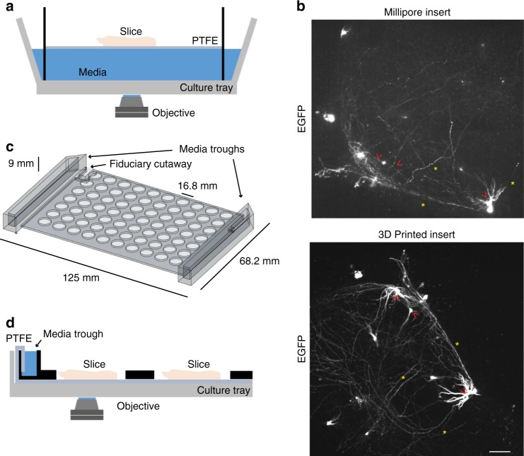
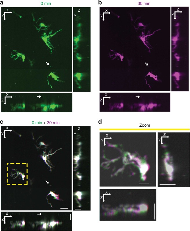
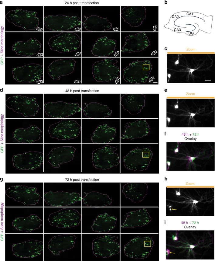
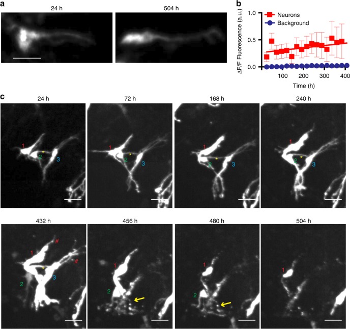
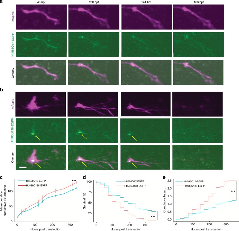
Current approaches for dynamic profiling of single cells rely on dissociated cultures, which lack important biological features existing in tissues. Organotypic slice cultures preserve aspects of structural and synaptic organisation within the brain and are amenable to microscopy, but established techniques are not well adapted for high throughput or longitudinal single cell analysis. Here we developed a custom-built, automated confocal imaging platform, with improved organotypic slice culture and maintenance. The approach enables fully automated image acquisition and four-dimensional tracking of morphological changes within individual cells in organotypic cultures from rodent and human primary tissues for at least 3 weeks. To validate this system, we analysed neurons expressing a disease-associated version of huntingtin (HTT586Q138-EGFP), and observed that they displayed hallmarks of Huntington's disease and died sooner than controls. By facilitating longitudinal single-cell analyses of neuronal physiology, our system bridges scales necessary to attain statistical power to detect developmental and disease phenotypes.

Keywords: Animal disease models; Confocal microscopy; Neurodegeneration; Time-lapse imaging; Tissue culture.


Conflict of interest statement

S.M.F. is the inventor of Robotic Microscopy Systems, U.S. Patent 7,139,415 and Automated Robotic Microscopy Systems, U.S. Patent Application 14/737,325, both assigned to The J. David Gladstone Institutes. The remaining authors declare no competing interests.

Figures

Fig. 1
Fig. 1
Optimisation of slice culture inserts for improved and high-throughput imaging organotypic tissue. a Cutaway illustration of a commercially available cell-culture insert (Millicell) in which a slice is suspended on a polytetrafluoroethylene (PTFE) membrane at the interface of the medium (blue) and air within a well of a culture tray. The inverted microscope objective images through medium to slice. b Comparison of representative confocal images of EGFP expression within transfected hippocampal slices imaged using Millicell insert (top) and 3D printed insert (bottom). Red arrowheads indicate comparable somata and yellow asterisks indicate neurites. Scale = 50 μm. c Illustration of the design of a 3D printed, full plate insert with attached medium troughs (arrows) sized to fit into standard cell culture tray. d Cutaway illustration of customised 3D printed array insert containing medium troughs (black objects) within a cell-culture tray, with reduced working distance of the microscope objective to the slice. PTFE membrane stretches along the length of the culture tray and wicks media from media trough to slice samples (pink objects)
Fig. 2
Fig. 2
Automated 4D imaging of single neurons within organotypic slice culture. a Maximum projections of representative hippocampal slice culture transfected with EGFP along XY (top left), XZ (bottom left) and YZ (top right) axes from automated confocal imaging with 184 z-slices. Arrow points to fluorescent beads placed between the transfected slice and the PTFE membrane. b A second confocal image stack of the same EGFP-transfected hippocampal slice culture taken after 30 min and projected into along XY (top left), XZ (bottom left) and YZ (top right) axes. c Alignment of 0-min (green) and 30-min (purple) 3D projections from a and b to create a 4D image projected along XY (top left), XZ (bottom left) and YZ (top right) axes. White areas indicate regions of overlap between time points, while areas of purple or green indicate regions of neuronal movement over time. XY scale = 50 μm, YZ and XZ scale = 20 μm. d Magnification of projections along XY (top left), XZ (bottom left) and YZ (top right) axes from yellow box in c showing subtle movements (areas of purple and green) over time within a single neuron in 4D. XY Scale = 15 μm, YZ and XZ scale = 20 μm
Fig. 3
Fig. 3
Longitudinal automated high-content, multiplexed imaging to detect changes within single neurons. a Maximum z-projections of 12 hippocampal slices transfected with EGFP and imaged with automation at high resolution 24 h post transfection (hpt) (left, scale bar 300 µm). Purple line represents edge of the slice. White cartoon inset shows the orientation of slice within the well. b Schematic of a hippocampal slice with locations of cornu ammonis (CA) 1, CA2, CA3, and dentate gyrus (DG) subregions. Miniaturised versions of same schematic were used in insets of a. c Magnification of three individual neurons within yellow box in a demonstrating high content of imaging approach. Scale = 50 μm. d Maximum z projections of the same 12 slices from a 48 hpt. e Expansion of same neurons within yellow box in c 48 hpt. f Overlay of imaging from 24 (magenta, same as c) and 48 hpt (green) showing differences in morphology over time. g Maximum z-projections of the same 12 slices from a and d 72 hpt. h Expanded view of yellow box in g. i Overlay of imaging 48 hpt (magenta, same as e) and 72 hpt (green) showing differences in morphology over time. Arrows indicate death of a neuron at 72 hpt that is present at 48 hpt
Fig. 4
Fig. 4
Complex changes in morphology preceding neuronal death revealed by longitudinal tracking of single cells in organotypic slice culture over weeks. a Maximum z-projections of a single EGFP-transfected neuron within a hippocampal slice culture imaged at 24 h post-transfection (hpt) (left) and 504 hpt (right) showing consistent EGFP signal. Scale = 40 μm. b Quantification of EGFP signal from single neurons (n = 12) imaged over 384 h showing increased EGFP signal over time. R2 = 0.8 SEMs are indicated. c Time-lapse time course of three neurons over 504 h showing changing morphology and neuronal death. Each neuron in a volume is labelled with a number (1–3) at the beginning of the experiment. When a cell dies, the software notes the event and the label disappears from the image. Yellow asterisks indicate apparent contacts between EGFP-labelled neurons. Red hashtags indicate areas of blebbing in unhealthy neurons. Yellow arrows indicate apparent neurite degeneration. Scale = 50 μm
Fig. 5
Fig. 5
Longitudinal imaging and tracking of single cells in human neocortical slice culture for extended time periods. a Maximum projections of longitudinal imaging of a representative GW19 human cortical slice expressing EGFP at 24, 27, 48, 72, 96, 120 and 144 hpi overlayed into a single image to show RG cell movement. Scale = 1 mm. b Zoom in of orange (top, cortical plate) and yellow (bottom, ventricular zone) boxes from a showing variable cell movement (white asterisks), process extensions (white arrowheads), and newly labelled cells (pink asterisks) over time across the slice. Scale = 100 µm. c (Left) Maximum projection of a representative GW22 slice across all Z and time series covering 0-468 hpi with 24 h intervals. (Left Inset) Zoom in of orange box showing region of furthest movement. Curved orange arrow indicates direction of 48° rotation to produce orange zoom in box. (Right) Automated cell tracking traces of the centroid from 719 segmented cells from left image overlayed across 469 h time course with each cell labelled in a distinct hue showing changes in its position over time. (Right Inset) Zoom in of orange box showing tracks of furthest movement. Curved orange arrow indicates direction of 48° rotation to produce orange zoom in box. Scale = 1 mm. d (Top) Quantification of number of cells segmented and tracked over time. Black line represents linear regression from which a rate of 10 cells per day was derived. (Middle) Quantification of mean cell area and (Bottom) mean distance of all cells tracked in c (R2 = 0.82, ANOVA Tukey multiple comparisons **p < 0.01, *p < 0.05). Green line represents linear regression of mean distance travelled from which an average velocity of −150 nm per day was derived (R2 = 0.6, ANOVA Tukey multiple comparisons **p < 0.01, *p < 0.05). Error bars show SD. e Longitudinal time series of an oRG cell from c (green arrow) undergoing divisions into daughter cells (yellow arrows), and serving as a scaffold for a migrating cell (red arrow) in xy from a single z slice (left panels, scale = 40 μm), zy brightest point projection (middle panels, scale = 30μm), and xz brightest point projection (right panels, scale = 40 μm) over the course of 432 h of imaging
Fig. 6
Fig. 6
Longitudinal imaging of HTT expressing neurons in organotypic slice culture. a, b Longitudinal imaging of single neurons from hippocampal slices co-transfected with the morphology marker mApple (top) and HTT586Q17-EGFP (a middle) or HTT586Q138-EGFP (b middle) showing morphological changes and death over the course of 168 h. Yellow arrows indicate putative inclusion body formation of HTT586Q138-EGFP. HTT586Q138-EGFP transfected neuron is dead at 168 hpt. c Plot of mean cumulative IB formation per slice over time, showing more small particles <80 μm2 accumulate by 336 hpt in slices transfected with HTT586Q138-EGFP (T-test ***p < 0.001) and accumulation proceeds at a faster rate (ANCOVA p < 0.05). d Neurons in slices transfected with HTT586Q17-EGFP survived longer than those transfected with HTT586Q138-EGFP. e Plot of the cumulative risk of survival of HTT586Q17-EGFP and HTT586Q138-EGFP showing linear increases in neuronal death over time. The hazard ratio of HTT586Q138-EGFP was 1.9 in comparison to httQ17-EGFP, indicating significantly increased toxicity of HTT586Q138-EGFP (Cox proportional hazard ***p < 0.001, 95% CI 1.5–2.5, HTT586Q17-EGFP n = 161 neurons, HTT586Q138-EGFP n = 180 neurons). SEMs are indicated. Scale bar = 20 μm

References

    1. Arrasate M, Mitra S, Schweitzer ES, Segal MR, Finkbeiner S. Inclusion body formation reduces levels of mutant huntingtin and the risk of neuronal death. Nature. 2004;431:805–810. doi: 10.1038/nature02998. - DOI - PubMed
    1. Barmada SJ, et al. Autophagy induction enhances TDP43 turnover and survival in neuronal ALS models. Nat. Chem. Biol. 2014;10:677–685. doi: 10.1038/nchembio.1563. - DOI - PMC - PubMed
    1. Finkbeiner S, Frumkin M, Kassner PD. Cell-based screening: extracting meaning from complex data. Neuron. 2015;86:160–174. doi: 10.1016/j.neuron.2015.02.023. - DOI - PMC - PubMed
    1. Hansen DV, Lui JH, Parker PR, Kriegstein AR. Neurogenic radial glia in the outer subventricular zone of human neocortex. Nature. 2010;464:554–561. doi: 10.1038/nature08845. - DOI - PubMed
    1. LaMonica BE, Lui JH, Hansen DV, Kriegstein AR. Mitotic spindle orientation predicts outer radial glial cell generation in human neocortex. Nat. Commun. 2013;4:1665. doi: 10.1038/ncomms2647. - DOI - PMC - PubMed

Publication types

MeSH terms