Review
. 2019 Feb 5;3(1):011501.
doi: 10.1063/1.5055873. eCollection 2019 Mar.

Modeling cardiac complexity: Advancements in myocardial models and analytical techniques for physiological investigation and therapeutic development in vitro

Review

Modeling cardiac complexity: Advancements in myocardial models and analytical techniques for physiological investigation and therapeutic development in vitro

Neal I Callaghan et al. APL Bioeng. .

Abstract

Cardiomyopathies, heart failure, and arrhythmias or conduction blockages impact millions of patients worldwide and are associated with marked increases in sudden cardiac death, decline in the quality of life, and the induction of secondary pathologies. These pathologies stem from dysfunction in the contractile or conductive properties of the cardiomyocyte, which as a result is a focus of fundamental investigation, drug discovery and therapeutic development, and tissue engineering. All of these foci require in vitro myocardial models and experimental techniques to probe the physiological functions of the cardiomyocyte. In this review, we provide a detailed exploration of different cell models, disease modeling strategies, and tissue constructs used from basic to translational research. Furthermore, we highlight recent advancements in imaging, electrophysiology, metabolic measurements, and mechanical and contractile characterization modalities that are advancing our understanding of cardiomyocyte physiology. With this review, we aim to both provide a biological framework for engineers contributing to the field and demonstrate the technical basis and limitations underlying physiological measurement modalities for biologists attempting to take advantage of these state-of-the-art techniques.


Figures

FIG. 1.
FIG. 1.
(a) An adult murine cardiomyocyte (CM), stained for sarcomeric α-actinin. Sarcomeres are continuous across the bundle of myofibrils that form the cell. Reprinted with permission from Ackers-Johnson et al., Circ. Res. 119(8), 909–920 (2016). Copyright 2016 Wolters Kluwer Health, Inc. (b) The cardiac action potential (top) is composed of four distinct phases, each with a specific ionic flux component (bottom). The cardiomyocyte produces a field potential (middle) that is highly correlated with the shape of its action potential. Reprinted with permission from Tertoolen et al., Biochem. Biophys. Res. Commun. 497(4), 1135–1141. Copyright 2018 Author(s), licensed under a Creative Commons Attribution 4.0 International License. (c) The resting membrane potential, and the precise shape of the action potential is specific to the CM subtype; nodal, atrial, and ventricular CMs have unique electrophysiological fingerprints. Reprinted with permission from Liu et al., Adv. Drug Delivery Rev. 96, 253–273. Copyright 2016 Elsevier. (d) The flux of contraction-enabling Ca2+ into the cytosol (black) is correlated with cell contraction strain (blue). Reprinted with permission from Ahola et al., Ann. Biomed. Eng. 46(1), 148–158. Copyright 2018 Author(s), licensed under a Creative Commons Attribution 4.0 International License. (e) Contractile CMs of different aspect ratios (3:1, 5:1, 7:1, unpatterned, and 1:1, respectively) deform compliant substrates allowing for the reconstruction of traction force vectors. Reprinted with permission from Ribeiro et al., Proc. Natl. Acad. Sci. 112(41), 12705–12710. Copyright 2015 National Academy of Sciences. (f) Transverse tubules responsible for initial Ca2+ inward flux (indicated by L-type Ca2+ channels, red) co-localize with sarcoplasmic reticulum (indicated by ryanodine receptor, green), which provides most of the cytosolic Ca2+ flux during a contraction. Reprinted with permission from Smyrnias et al., Cell Calcium 47(3), 210–223 (2010). Copyright 2010 Elsevier. (g) Gap junctions establish a functional syncytium, allowing HTTS (hydroxypyrene-1,3,6-trisulfonic acid, trisodium salt) dye propagation between cells. Knockdown of intercalated disc trafficking protein Tmem65 ablates gap junction formation. Reprinted by permission from Sharma et al., Nat. Commun. 6, 8391 (2015). Copyright Springer Nature.
FIG. 2.
FIG. 2.
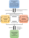
The role of advancing techniques to assess the cardiomyocyte function in vitro in the workflow to improved pharmaceutical testing and clinical outcomes. Both hypothesis- and discovery-driven research studies push each other forward, while informing iterations of cell and tissue models and devices, modalities, and techniques used in experimentation. The discoveries from fundamental science can then be translated to the clinic, while clinical findings can be used as a starting point for further fundamental inquiry.
FIG. 3.
FIG. 3.
Negative correlation between the current usability of representative CM models and their final utility in advancing the field as a function of their ability to recapitulate complex and emergent CM physiological phenomena such as electrophysiology, contractility, signaling, and metabolism. By using improved functional characterization, which fuels improved experimentation, both the ease of use and scientific value and translatability can be increased.
FIG. 4.
FIG. 4.
(a) Common principles underlying most measurements of contraction; an axial force can be measured directly using an isometric or strain gauge force transducer. Conversely, during an isotonic contraction, perpendicular cell thickening can be used as a correlative measurement due to the conservation of volume within a contracting cell. The titin structure within the CM offers a baseline of passive resistance (Tpassive), to which the applied force vector from active contraction (Fa) is added. (b) Principles of contraction within a sarcomere of stable A-band dimensions that allow for sarcomeric shortening measurements of isotonic (above) and isometric contraction (below) such as seen in TEM studies. The scale bar on the left is 1 μm. (c) The heart makes use of both near-isotonic (auxotonic) and isometric contractions with each cycle, as illustrated by a standard pressure-volume loop. The cycle occurs in a counter-clockwise direction, with systole and diastole ending before isometric phases at the top-left and bottom-right corners of the loop, respectively. Passive diastolic filling at near-constant pressure results in eccentric contraction in the myocardium, which corresponds to the degree of preload or end-diastolic pressure.

References

    1. Benjamin E. J., Virani S. S., Callaway C. W., Chamberlain A. M., Chang A. R., Cheng S., Chiuve S. E., Cushman M., Delling F. N., Deo R., de Ferranti S. D., Ferguson J. F., Fornage M., Gillespie C., Isasi C. R., Jiménez M. C., Jordan L. C., Judd S. E., Lackland D., Lichtman J. H., Lisabeth L., Liu S., Longenecker C. T., Lutsey P. L., Mackey J. S., Matchar D. B., Matsushita K., Mussolino M. E., Nasir K., O'Flaherty M., Palaniappan L. P., Pandey A., Pandey D. K., Reeves M. J., Ritchey M. D., Rodriguez C. J., Roth G. A., Rosamond W. D., Sampson U. K. A., Satou G. M., Shah S. H., Spartano N. L., Tirschwell D. L., Tsao C. W., Voeks J. H., Willey J. Z., Wilkins J. T., Wu J. H., Alger H. M., Wong S. S., and Muntner P., Circulation 137, e67 (2018).10.1161/CIR.0000000000000558 - DOI - PubMed
    1. Qu Z. and Weiss J. N., Annu. Rev. Physiol. 77, 29 (2015).10.1146/annurev-physiol-021014-071622 - DOI - PMC - PubMed
    1. Schotten U., Dobrev D., Platonov P. G., Kottkamp H., and Hindricks G., J. Intern. Med. 279, 428 (2016).10.1111/joim.12492 - DOI - PubMed
    1. Nattel S. and Dobrev D., Nat. Rev. Cardiol. 13, 575 (2016).10.1038/nrcardio.2016.118 - DOI - PubMed
    1. Zhou P. and Pu W. T., Circ. Res. 118, 368 (2016).10.1161/CIRCRESAHA.116.308139 - DOI - PMC - PubMed