Review
. 2019 Dec;22(4):423-431.
doi: 10.1007/s40477-019-00379-0. Epub 2019 May 8.

Skin cancer: findings and role of high-resolution ultrasound

Affiliations
Review

Skin cancer: findings and role of high-resolution ultrasound

Orlando Catalano et al. J Ultrasound. 2019 Dec.

Abstract

Currently available high-resolution transducers allow a detailed ultrasound (US) assessment of skin tumors. US complements clinical examination, dermoscopy, and biopsy in the initial differential diagnosis, surgical planning, locoregional staging, and follow-up of patients with skin malignancies. It is important for dermatologists, skin surgeons, and US operators to be aware of the US imaging findings and to recognize the clinical scenarios where imaging is indicated in the management of skin cancer. The purpose of this review article is to address the most common indications for US in skin oncology and to provide a comprehensive guide to the gray-scale and color-Doppler findings in cutaneous malignant tumors.

Keywords: Dermatology ultrasound; Melanoma; Skin tumors; Skin ultrasound; Soft tissues.


Conflict of interest statement

The authors declare that they have no conflict of interest.

Figures

Fig. 1
Fig. 1
Approximate prevalence of skin cancers. BCC basal cell carcinoma, SCC squamous cell carcinoma, CM cutaneous melanoma
Fig. 2
Fig. 2
Basal cell carcinoma of the nasion. Patient photograph (a). Ultrasound scan with tumor thickness measurement (b). The lesion is hypoechoic and relatively homogeneous, with some hyperechoic spots. It is adherent to the bony cortex
Fig. 3
Fig. 3
Ulcerated basal cell carcinoma of the nose ala. Patient photograph (a). Ultrasound scan (b). The lesion is hypoechoic and relatively homogeneous, with some hyperechoic spots. The tumor does not infiltrate the cartilage (calipers)
Fig. 4
Fig. 4
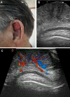
Ear scapha squamous cell carcinoma. Patient photograph (a). Ultrasound scan (b). The 7-mm thick lesion (calipers) is hypoechoic and relatively homogeneous. There is no infiltration of the cartilage. Color-Doppler scan (c). Discrete vascularization, with vessels mostly arranged vertically, starting from two deep vascular poles
Fig. 5
Fig. 5
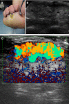
Sole melanoma. Patient photograph (a). Ultrasound scan (b). The 3-mm thick tumor is hypoechoic and homogeneous. Directional power-Doppler scan (c). Very intense tumor vascularization
Fig. 6
Fig. 6
Dermatofibrosarcoma protuberans of the head (frontal region), local recurrence. Ultrasound scan (a). Thick, homogeneous, and ill-defined lesion, adherent to the bone surface. Directional power-Doppler scan (b). Strong, irregular vascularization at power-Doppler imaging
Fig. 7
Fig. 7
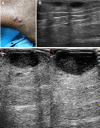
Thigh metastases from Kaposi sarcoma. Patient photograph (a). Ultrasound scans, taken at 13.5 MHz (b) and at 22 MHz (c). The latter image shows the two hypodermis, hypoechoic nodules with a superior detail compared to the former

References

    1. Bard RL. High-frequency ultrasound examination in the diagnosis of skin cancer. Dermatol Clin. 2017;35:505–511. doi: 10.1016/j.det.2017.06.011. - DOI - PubMed
    1. Que SKT, Zwald FO, Schmults CD. Cutaneous squamous cell carcinoma: incidence, risk factors, diagnosis, and staging. J Am Acad Dermatol. 2018;78:237–247. doi: 10.1016/j.jaad.2017.08.059. - DOI - PubMed
    1. Wortsman X, Alfageme F, Roustan G, Arias-Santiago S, Martorell A, Catalano O, Scotto di Santolo M, Zarchi K, Bouer M, Gonzalez C, Bard R, Mandava A, Gaitini D. Guidelines for performing dermatologic ultrasound examinations by the DERMUS Group. J Ultrasound Med. 2016;35:577–580. doi: 10.7863/ultra.15.06046. - DOI - PubMed
    1. Alfageme Roldán F. Handbook of skin ultrasound. Charleston: CreateSpace Independent Publishing Platform; 2013.
    1. Alfageme Roldán F. Ultrasound skin imaging. Actas Dermosifiliogr. 2014;105:891–899. doi: 10.1016/j.ad.2013.11.015. - DOI - PubMed