. 2019 Jun 19;11(6):1128-1139.
doi: 10.1039/c9mt00057g.

Single nucleotide polymorphisms in the human ATP7B gene modify the properties of the ATP7B protein

Affiliations

Single nucleotide polymorphisms in the human ATP7B gene modify the properties of the ATP7B protein

Courtney J McCann et al. Metallomics. .

Erratum in

Abstract

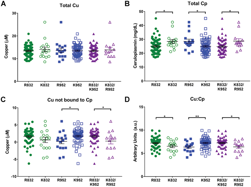
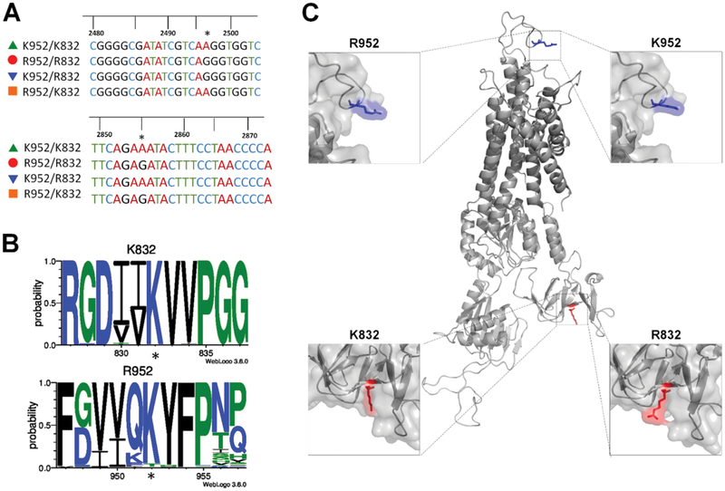
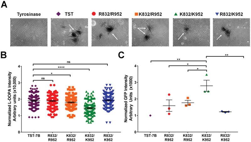
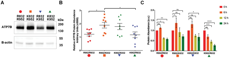
Single nucleotide polymorphisms (SNPs) are the largest source of sequence variation in the human genome. However, their functional significance is not well understood. We show that SNPs in the Wilson disease gene, ATP7B, that produce amino-acid substitutions K832R and R952K, modulate ATP7B properties in vitro and influence serum copper (Cu) status in vivo. The presence of R832 is associated with a lower ATP7B abundance and a diminished trafficking in response to elevated Cu. The K832R substitution alters surface exposure of amino acid residues in the actuator domain and increases its conformational flexibility. All SNP-related ATP7B variants (R832/R952, R832/K952, K832/K952, and K832/R952) have Cu-transport activity. However, the activity of ATP7B-K832/K952 is lower compared to other variants. In humans, the presence of K952 is associated with a higher fraction of exchangeable Cu in serum. Thus, SNPs may modulate the properties of ATP7B and the organism Cu status.


Conflict of interest statement

CONFLICTS OF INTEREST

Dr. R. Squitti is a consultant Canox4drug SPA (Italy) and Igea Research Inc. (Miami, FL). Other authors have no conflicts of interest to declare.

Figures

Fig. 1
Fig. 1
Presence of R832 and K952 correlates with changes in serum Cu markers. (A) Total serum Cu concentration (mM) in healthy individuals with indicated genotypes. (B) Serum Cp (mg dL1) from healthy individuals with indicated genotypes; (C) concentration of non-Cp bound Cu in the serum of healthy individuals with indicated genotypes. (D) Ratio of total Cu to total Cp in the serum of healthy individuals with indicated genotypes. Normal, healthy levels range from 16–24 mM for Cu [3], 20–40 mg dL1 for Cp [3], 0.07–2.19 mM for non-Cp Cu [4], and 6.8 for Cu : Cp [4]. n = 16 for R832, n = 65 for K832, n = 16 for R952, n = 65 for K952, n = 13 for K832/R952, and n = 62 for R832/K952. Values are reported as means SEM. Significance was determined by Student’s t-test; *p o 0.05 and **p o 0.01.
Fig. 2
Fig. 2
Location, conservation, and potential impact of SNP-related substitutions on ATP7B structure (A) sequence of four generated ATP7B plasmids shows either an adenine (encoding Lys) or a guanine (encoding Arg) at positions 2495 and 2855, marked by asterisks. (B) Conservation logos of 11-AA sequences centered on K832 or R952 were generated using 165 animal ATP7B orthologues. The SNP related AA are marked with asterisks. K832 is well-conserved among ATP7B homologs, whereas R952 is specific for human ATP7B. (C) Homology model of ATP7B based on the crystal structure of L. pneumophila CopA [1]. Top panels illustrate location of the R/K variants at position 952. Bottompanels show location of the R/K variants at position 832 in the A-domain of ATP7B.
Fig. 3
Fig. 3
ATP7B-K952/K832 has a lower Cu-transport activity compared to other variants. (A) YST cells were transfected with a plasmid expressing pTyrosinase (pTyr) alone (Tyrosinase) or co-transfected with pTyr and twin strep-tagged ATP7B (TST-ATP7B) or pTyr and one of the GFP-tagged ATP7B variants. The Cu transport activity of all ATP7B variants was evaluated by activation of tyrosinase (dark brown reaction product, marked by arrows, indicates tyrosinase activity). (B) Quantitation of tyrosinase activity. Pigment intensity is normalized to pigment area. (~) TST-ATP7B in purple, (K) R832/ R952 in red, (‘) K832/R952 in orange, (m) K832/K952 in green, and (.) R832/K952 in blue. n = 4, over 40 areas counted per n. (C) Quantitation of ATP7BGFP levels in cells used in the Cu-transport assay. GFP fluorescence intensity was normalized to the area, the fluorescence intensity of control cells expressing pTyr only, and finally to the fluorescent intensity of cells expressing TST-ATP7B, which was taken as 1. n = 3. All values are reported as means SEM. Significance was determined by one-way ANOVA with Fisher’s LSD test; *p o 0.05, **p o 0.01, and ****p o 0.0001.
Fig. 4
Fig. 4
Presence of R832 is associated with lower ATP7B abundance. (A) HEK293A cells were transfected with ATP7B-GFP variants for 20 h, whole cell lysates were prepared and run on SDS-PAGE (20 mg of protein per lane).Western blot shows the relative abundance of each ATP7B variant. b-Actin is used as a loading control. (B) Densitometry of Western blots, n = 8. (K) R832/R952 in red, (‘) K832/R952 in orange, (.) R832/K952 in blue, and (m) K832/K952 in green. Intensities of ATP7B bands were normalized to b-actin and plotted. Means SEM are indicated by horizontal lines. Significance was determined by one-way ANOVA with Fisher’s LSD test; *po0.05. (C) The R832-containing ATP7B variants aremore susceptible to degradation than K832 variants. HEK293A cells were transfected, as described above, and then treated with 50 mM cycloheximide (CHX) for 0, 6, 12, or 24 h. For each time point,whole cell lysateswere prepared and equal amounts of total protein (5 mg per lane) were run on SDS-PAGE; ATP7B was visualized by Western blotting. The bars indicate the intensity of ATP7B bands at 6 h (orange), 12 h (yellow), and 24 h (green) relative to intensity at 0 time point (red) which is taken as 1. n = 4. Values are reported as means SEM. Significance was determined by one-way ANOVA with Fisher’s LSD test; *p o 0.05, **p o 0.01, ***p o 0.001, and ****p o 0.0001.
Fig. 5
Fig. 5
Localization of ATP7B variants in HEK293 cells in low and high copper HEK293 cells were transfected with one of the indicated ATP7B-GFP variant (green), cells were treated for 4 h with either 25 mM TTM (top panels) or 100 mM CuSO4 (bottom panels) and immunostained for trans Golgi network marker TGN46 (red). Colocolization is indicated by yellow; the loss of colocalization is indicative of ATP7B trafficking (n = 4).
Fig. 6
Fig. 6
R832 increases A-domain flexibility (A) surface electrostatics of the ATP7B A-domain [2] containing R832/K832 variants quantified using AMBER (left pair) or PARSE (right pair). (B) R832 increases the conformational flexibility of the A-domain. Gaussian network mode-based normal mode frequencies of (‘) K832 in black and (K) R832 in red (C) the radius of gyration of the A-domain containing either K832 (K, red) or R832 (‘, gray). (D) The variation of SASA values for AA residues in b sheet 3 during 200 ns simulation: R832 (red, filled circles), K832 (red, open circles): V833 (K, blue), V834 (yellow), I830 (green) and V831 (magenta). (E) Variation in SASA values for residues from the TGE motif in the R832 and K832 A domains: T858 (green), E860 (magenta), and G859 (blue). (F) Cross-correlation maps of AA motions in K832 (top) and R832 (bottom) A-domains during 200 ns simulation. Positive values (yellow) indicate motion in the same direction, whereas negative values (blue) indicate motion in the opposite direction. All values are reported as means SEM. Significance was determined by unpaired Student’s t-test; **p o 0.01, ****p o 0.0001.

References

    1. Lutsenko S, Copper trafficking to the secretory pathway, Metallomics, 2016, 8, 840–852. - PMC - PubMed
    1. van den Berghe PV and Klomp LW, New developments in the regulation of intestinal copper absorption, Nutr Rev, 2009, 67, 658–672. - PubMed
    1. Michalczyk AA, Rieger J, Allen KJ, Mercer JF and Ackland ML, Defective localization of the Wilson disease protein (ATP7B) in the mammary gland of the toxic milk mouse and the effects of copper supplementation, Biochem J, 2000, 352 Pt 2, 565–571. - PMC - PubMed
    1. Braiterman LT, Gupta A, Chaerkady R, Cole RN and Hubbard AL, Communication between the N and C termini is required for copper-stimulated Ser/Thr phosphorylation of Cu(I)-ATPase (ATP7B), J Biol Chem, 2015, 290, 8803–8819. - PMC - PubMed
    1. Lutsenko S, Barnes NL, Bartee MY and Dmitriev OY, Function and regulation of human copper-transporting ATPases, Physiol Rev, 2007, 87, 1011–1046. - PubMed

Publication types

MeSH terms

LinkOut - more resources