. 2019 Nov;70(5):1674-1689.
doi: 10.1002/hep.30706. Epub 2019 Jun 22.

Proteasomal Degradation of Enhancer of Zeste Homologue 2 in Cholangiocytes Promotes Biliary Fibrosis

Affiliations

Proteasomal Degradation of Enhancer of Zeste Homologue 2 in Cholangiocytes Promotes Biliary Fibrosis

Nidhi Jalan-Sakrikar et al. Hepatology. 2019 Nov.

Abstract

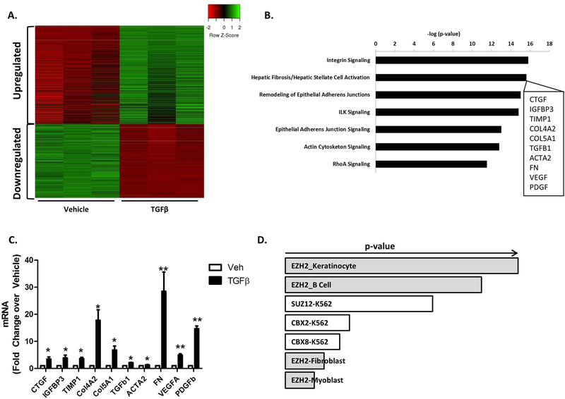
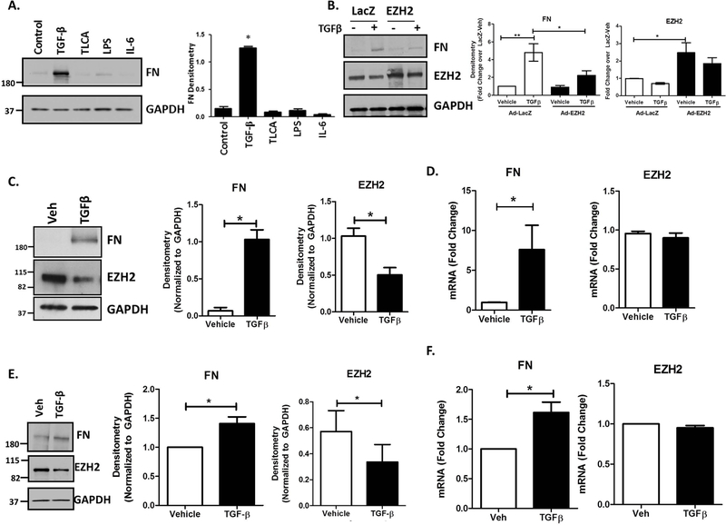
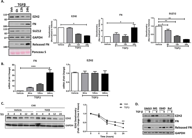
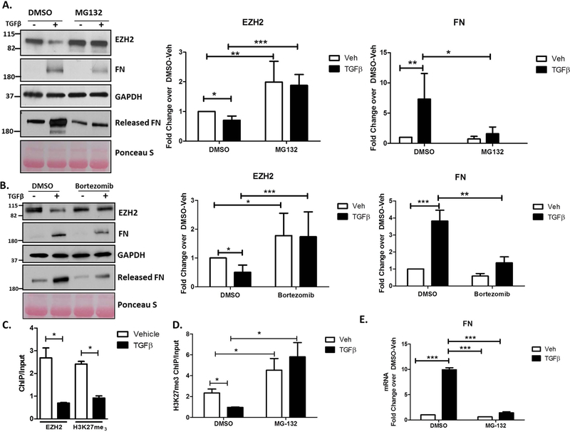
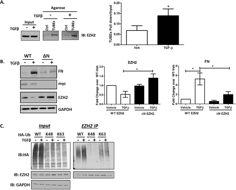
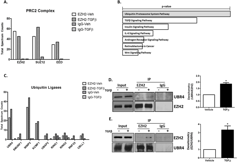
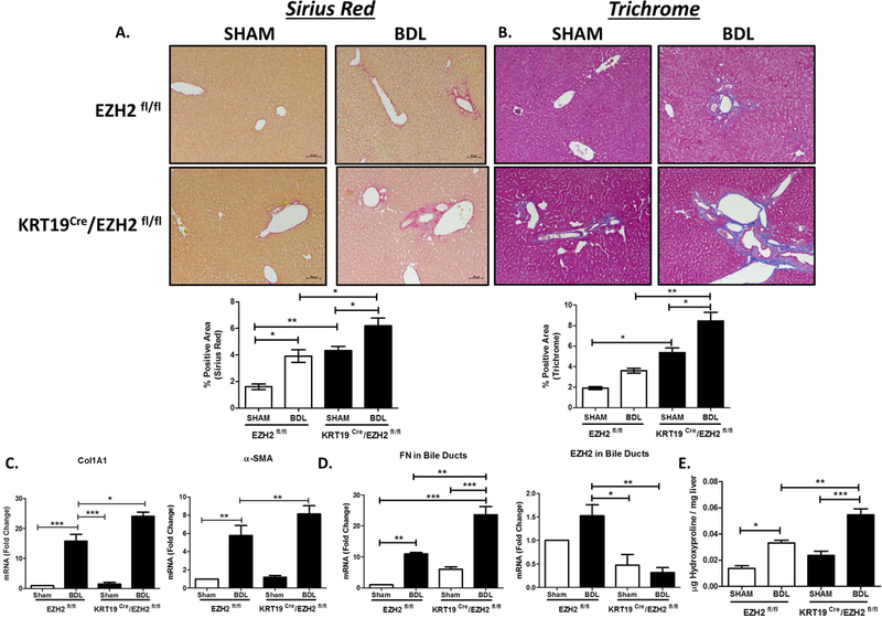
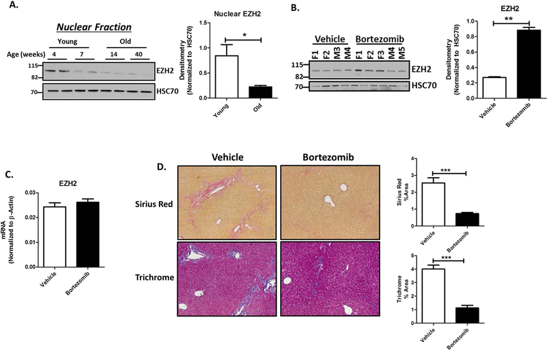
During biliary disease, cholangiocytes become activated by various pathological stimuli, including transforming growth factor β (TGF-β). The result is an epigenetically regulated transcriptional program leading to a pro-fibrogenic microenvironment, activation of hepatic stellate cells (HSCs), and progression of biliary fibrosis. This study evaluated how TGF-β signaling intersects with epigenetic machinery in cholangiocytes to support fibrogenic gene transcription. We performed RNA sequencing in cholangiocytes with or without TGF-β. Ingenuity pathway analysis identified "HSC Activation" as the highly up-regulated pathway, including overexpression of fibronectin 1 (FN), connective tissue growth factor, and other genes. Bioinformatics identified enhancer of zeste homologue 2 (EZH2) as an epigenetic regulator of the cholangiocyte TGF-β response. EZH2 overexpression suppressed TGF-β-induced FN protein in vitro, suggesting FN as a direct target of EZH2-based repression. Chromatin immunoprecipitation assays identified an FN promoter element in which EZH2-mediated tri-methylation of lysine 27 on histone 3 is diminished by TGF-β. TGF-β also caused a 50% reduction in EZH2 protein levels. Proteasome inhibition rescued EZH2 protein and led to reduced FN production. Immunoprecipitation followed by mass spectrometry identified ubiquitin protein ligase E3 component N-recognin 4 in complex with EZH2, which was validated by western blotting in vitro. Ubiquitin mutation studies suggested K63-based ubiquitin linkage and chain elongation on EZH2 in response to TGF-β. A deletion mutant of EZH2, lacking its N-terminal domain, abrogates both TGF-β-stimulated EZH2 degradation and FN release. In vivo, cholangiocyte-selective knockout of EZH2 exacerbates bile duct ligation-induced fibrosis whereas MDR2-/- mice are protected from fibrosis by the proteasome inhibitor bortezomib. Conclusion: TGF-β regulates proteasomal degradation of EZH2 through N-terminal, K63-linked ubiquitination in cholangiocytes and activates transcription of a fibrogenic gene program that supports biliary fibrosis.


Figures

Figure 1
Figure 1
TGFβ Drives Expression of an HSC-Activating Gene Program in Cholangiocytes. A. Heat Map representing the gene changes in a cholangiocytes cell line (HIBEC) treated with 10ng/ml TGFβ for 48 hours (green: upregulated gene set; red: downregulated gene set). n=3 B. Ingenuity Pathway Analysis (IPA) shows Integrin signaling and Hepatic Fibrosis/Hepatic Stellate Cell Activation as the most significantly upregulated pathways (inset shows 10 representative genes from the Heaptic Stellate Cell Activation Pathway). C. RT-PCR in cholangiocytes treated with 10ng/ml TGFβ for 24 hours confirms upregulation of HSC activation genes. *p<0.01 and **p<0.001. All error bars are SEM, n=3 D. EnrichR analysis identified EZH2 as an epigenetic regulator of the cholangiocyte TGFβ response (p-vaue represents Fisher exact test).
Figure 2
Figure 2
Cholangiocytes Upregulate FN in Response to TGFβ. A. H69 cells treated for 24 hours with several agonists prominent in biliary disease show that only TGFβ stimulates FN production. TGFβ: 10ng/ml, TLCA: 20uM, LPS: 200ng/ml, IL-6: 50ng/ml. Western blot for FN and EZH2 with GAPDH as a loading control. These expression levels were quantified by densitometry (right). *p<0.01. All error bars are SEM, n=3. B. Western blot showing repression of TGFβ–stimulated FN by overexpression of EZH2 using adenoviral constructs (AdEZH2) (left). Quantification of the blot for EZH2 and FN normalized to LacZ-vehicle. *p<0.01 and **p<0.001. All error bars are SEM, n=4. C. TGFβ treatment for 24 hours stimulates FN production in cholangiocytes and a reduction in total EZH2 levels. Immunoblotting for FN and EZH2 on H69 cells treated with TGFβ for 24 hours (left). These were quantified by densitometry for FN and EZH2 (right) *p<0.01 compared to vehicle. All error bars are SEM, n=3. D. RT-PCR analysis on H69 cells treated with TGFβ for 24 hours shows an increase in FN gene expression with no change in EZH2 gene expression. *p<0.01 compared to vehicle. All error bars are SEM, n=3. E. Cholangiocytes isolated from WT miee were treated with 10ng/ml TGFβ for 24 hours. Western blotting for FN and EZH2 show TGFβ -stimulated FN production with a concurrent decrease in EZH2 protein (left). Densitometry from 3 different experiments shows an increase in FN and a decrease in EZH2 protein (right). *p<0.01 compared to vehicle. All error bars are SEM, n=3. F. RT-PCR analysis on mouse cholangiocytes treated with TGFβ shows an approximately 2-fold increase in FN gene transcription with no change in EZH2 gene transcription. *p<0.01 compared to vehicle. All error bars are SEM, n=3.
Figure 3
Figure 3
TGFβ Stimulation Leads to EZH2 Protein Degradation in Cholangiocytes. A. Western blotting on H69 cells treated with TGFβ for 6, 12, and 24 hours show a time-dependent increase in FN production as well as its release into the media. This was seen in conjunction with a decrease in EZH2 and SUZ12 protein at 12 hours with a 50% decrease at 24 hours (left). Densitometry for FN, EZH2 and SUZ12 show statistically significant differences compared to vehicle at the 24 hour time point. * p<0.01. All error bars are SEM, n=3. B. RT-PCR analysis for FN and EZH2 gene transcription in H69 cells treated with TGFβ. FN gene transcription increases with time after TGFβ treatment with no change in EZH2 gene transcription. ***p<0.0001. All error bars are SEM, n=3. C. Western blot for EZH2 in cholangiocytes treated with cycloheximide (CHX: 40µg/ml) for the indicated time (hours) in the presence of vehicle or TGFβ (left). Right shows the rate of EZH2 protein turnover with CHX in the presence of vehicle or TGFβ. **p<0.001. All error bars are SEM, n=4 D. Western blotting on lysates of H69 cells treated with vehicle or TGFβ in presence of various inhibitors (MG-132, E64D, and Bafilomycin with DMSO as control). Only inclusionof MG-132 rescued EZH2 levels with concurrent decrease in FN levels.
Figure 4
Figure 4
EZH2 Degradation Occurs via the Ubiquitin Proteasome System Downstream of TGFβ. A. The lysates of H69 cells treated with vehicle or TGFβ in the presence of DMSO or MG-132 were blotted for EZH2, FN, and GAPDH (loading control) and the cell media was blotted for released FN with Ponceau Stain as a loading control for the media (left). Densitometry for EZH2 and FN show rescue of EZH2 levels in presence of MG-132 and concomitant decrease in FN production (right). * p<0.01, **p<0.001, ***p<0.0001. All error bars are SEM, n=4. B. Protein lysates from H69 cells treated with vehicle or TGFβ in the presence of the clinically-relevent proteasome inhibitor, Bortezomib or DMSO were immunoblotted for EZH2, FN, and GAPDH (loading control) and the cell media was blotted for released FN with Ponceau Stain as loading control for the media (left). Densitometric quantification reveals recue of EZH2 protein levels with Bortezomib treatment and a concurrent decrease in FN levels (right). * p<0.01, **p<0.001, ***p<0.0001. All error bars are SEM, n=3. C. Cells treated with vehicle or TGFβ were analyzed by ChIP for the H3K27me3 mark and EZH2 occupancy on the FN promoter. The assay demonstrates reduced levels of the H3K27me3 mark consistent with reduced EZH2 occupancy on the FN promoter. * p<0.01. All error bars are SEM, n=3. D. H69 treated with vehicle or TGFβ in the presence of DMSO or MG-132 were analyzed by ChIP for the H3K27me3 mark on the FN promoter. MG-132 rescues TGFβ-mediated reduction in H3K27me3. * p<0.01. All error bars are SEM, n=3. E. RT-PCR analysis for FN on H69 cells treated with vehicle or TGFβ in the presence of DMSO or MG-132. The TGFβ-mediated increase in FN gene transcription is abrogated in the presence of MG-132. ***p<0.0001. All error bars are SEM, n=3.
Figure 5
Figure 5
The N-Terminus of EZH2 is Poly-Ubiquitinated by Through K63-Based Ubiquitin Linkage. A. Lysates of vehicle or TGFβ treated H69 cells were incubated with control (Ctrl) or TUBEs agarose beads. Immunoblotting for EZH2 on the beads reveals increased EZH2 pull-down in the presence of TGFβ (left). Densitometry on the right show a 1.5-fold increase in EZH2 binding to TUBEs agarose in presence of TGFβ. * p<0.01. All error bars are SEM, n=3. B. H69 cells were transfected with N-terminal, myc-tagged, full-length EZH2 (WT) or N-terminal deletion mutant of EZH2 (ΔN). Lysates of transfected cells treated with vehicle or TGFβ were immunoblotted for FN, myc, EZH2, and GAPDH (loading control). Densitometry reveals that TGFβ-induced loss of WT EZH2 protein was abolished with deletion of the N-terminal 100 residues, which further blocked TGFβ-stimulated FN production. * p<0.01. All error bars are SEM, n=3. C. H69 cells transfected with WT, K48, or K63 HA-Ubiquitin (Ub) were treated with either vehicle or TGFβ. Lysates were immunoprecipitated with EZH2 antibody and immunoblotted for HA and EZH2. HA blot on immunoprecipitates demonstrate EZH2 ubiquitination with WT HA-Ub and K63 HA-Ub.
Figure 6
Figure 6
EZH2 is Ubiquitinated by the E3 Ubiquitin Ligase, UBR4. A. Total Spectrum Counts of PRC2 complex components; EZH2, EED, and SUZ12 identified by mass spectrometry in EZH2 and IgG immunoprecipitates from vehicle and TGFβ treated H69 cells. B. EnrichR analysis on proteins co-immunoprecipitated with EZH2 identified the Ubiquitin Proteasome System and TGFβ Signaling as top pathways (p-vaue represents Fisher exact test). C. Total Spectrum Counts of the Ubiquitin ligases co-immunoprecipitated with EZH2 and IgG (control) from H69 cells treated with vehicle or TGFβ. UBR4: Ubiquitin Protein Ligase E3 Component N-Recognin 4, SMURF1: SMAD Specific E3 Ubiquitin Protein Ligase 1, WWP2: WW Domain Containing E3 Ubiquitin Protein Ligase 2, KCMF1: Potassium Channel Modulatory Factor 1, UBAP2: Ubiquitin Associated Protein 2, RING1: Ring Finger Protein 1, RING2: Ring Finger Protein 2, RNF25: Ring Finger Protein 25, CBLL1: Cbl Proto-Oncogene Like 1. D. Western blotting on EZH2 and IgG immunoprecipitates from H69 cells treated with vehicle or TGFβ confirms the presence of UBR4 (left). Densitometry on the right shows a1.6-fold increase in UBR4 co-immunoprecipitated with EZH2. *p<0.01. All error bars are SEM, n=4. E. Western blotting on UBR4 and IgG immunoprecipitates from H69 cells treated with vehicle or TGFβ confirms EZH2 in complex with UBR4 (left). Ddensitometric quantification demonstrates a 3-fold increase in EZH2 co-immunoprecipitated with UBR4 after TGF β. *p<0.01. All error bars are SEM, n=4.
Figure 7
Figure 7
Cholangiocyte-Selective Knockout of EZH2 Exacerbates Biliary Fibrosis. A. Liver histology shows Sirius red staining on EZH2fl/fl and KRT19CRE/EZH2fl/fl animals with sham or BDL surgery (Top). Bottom shows quantification of % positive area for Sirius red (n=4–5 animals per group). *p<0.01, **p<0.001 All error bars are SEM. B. Liver histology showing Trichrome staining on EZH2fl/fl and KRT19CRe/EZH2fl/fl animals with sham or BDL surgery (Top). Bottom shows quantification of % positive area for Trichrome (n=4–5 animals per group). *p<0.01, **p<0.001. All error bars are SEM. C. RT-PCR analysis for collagen1A1 and alpha-SMA gene expression in whole liver tissue from EZH2fl/fl and KRT19 Cre/EZH2fl/fl animals with Sham and BDL surgery (n=4–5 animals per group). *p<0.01, **p<0.001, ***p<0.0001. All error bars are SEM. D. RT-PCR analysis for FN1 and EZH2 in isolated bile ducts using LCM from EZH2fl/fl and KRT19 Cre/EZH2fl/fl animals with Sham and BDL surgery (n=4–5 animals per group). *p<0.01, **p<0.001, ***p<0.0001. All error bars are SEM. E. Hydroxyproline analysis on liver tissue from EZH2 EZH2fl/fl and KRT19 Cre/EZH2fl/fl animals with Sham and BDL surgery (n=4–5 animals per group). *p<0.01, **p<0.001, ***p<0.0001. All error bars are SEM.
Figure 8
Figure 8
Proteasome Inhibition Restores EZH2 Protein and Reduces Fibrosis in Mdr−/− Mice. A. Western blotting on nuclear fractions of liver lysates from young (4 and 7 weeks) and old (14 and 40 weeks) Mdr2−/− mice show decreasing levels of EZH2 protein (left) with HSC70 as a loading control. Densitometry on the right shows a 3-fold decrease in EZH2 levels. *p<0.01. All error bars are SEM, n=4. B. Mdr2−/− mice were treated with 1mg/kg Bortezomib from 7weeks to 11 weeks of age (males and females). Immunoblotting for EZH2 on nuclear fraction of liver lysates from both vehicle and Bortezomib treated mice show an increase in EZH2 protein levels with Bortezomib treatment (left), F: female, M: Male. Densitometry on the right shows 3-fold increase EZH2 protein when normalized to HSC70 as loading control. **p<0.001. All error bars are SEM, n=3. C. RT-PCR analysis on the liver tissue from vehicle and Bortezomib treated Mdr2−/− mice show no change in EZH2 gene expression. All error bars are SEM, n=3. D. Liver histology from Mdr2−/− mice treated with vehicle or Bortezomib show reduced fibrosis by Sirius red and Trichrome staining (left). Right shows the quantification for the % positive area for Sirius red and trichrome staining. ***p<0.0001. All error bars are SEM, n=4–5 animals per group.

References

    1. Abshagen K, Konig M, Hoppe A, Muller I, Ebert M, Weng H, Holzhutter HG, et al. Pathobiochemical signatures of cholestatic liver disease in bile duct ligated mice. BMC Syst Biol 2015;9:83. - PMC - PubMed
    1. Fabris L, Spirli C, Cadamuro M, Fiorotto R, Strazzabosco M. Emerging Concepts in Biliary Repair and Fibrosis. Am J Physiol Gastrointest Liver Physiol 2017:ajpgi 00452 02016. - PMC - PubMed
    1. Lazaridis KN, LaRusso NF. Primary Sclerosing Cholangitis. N Engl J Med 2016;375:2501–2502. - PubMed
    1. Jones H, Hargrove L, Kennedy L, Meng F, Graf-Eaton A, Owens J, Alpini G, et al. Inhibition of mast cell-secreted histamine decreases biliary proliferation and fibrosis in primary sclerosing cholangitis Mdr2(−/−) mice. Hepatology 2016;64:1202–1216. - PMC - PubMed
    1. Pi L, Robinson PM, Jorgensen M, Oh SH, Brown AR, Weinreb PH, Trinh TL, et al. Connective tissue growth factor and integrin alphavbeta6: a new pair of regulators critical for ductular reaction and biliary fibrosis in mice. Hepatology 2015;61:678–691. - PMC - PubMed

Publication types

Substances