. 2019 Jun 10;20(6):2327-2337.
doi: 10.1021/acs.biomac.9b00304. Epub 2019 May 29.

Nano- and Micropatterned Polycaprolactone Cellulose Composite Surfaces with Tunable Protein Adsorption, Fibrin Clot Formation, and Endothelial Cellular Response

Affiliations

Nano- and Micropatterned Polycaprolactone Cellulose Composite Surfaces with Tunable Protein Adsorption, Fibrin Clot Formation, and Endothelial Cellular Response

Tamilselvan Mohan et al. Biomacromolecules. .

Abstract

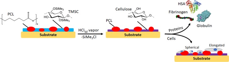
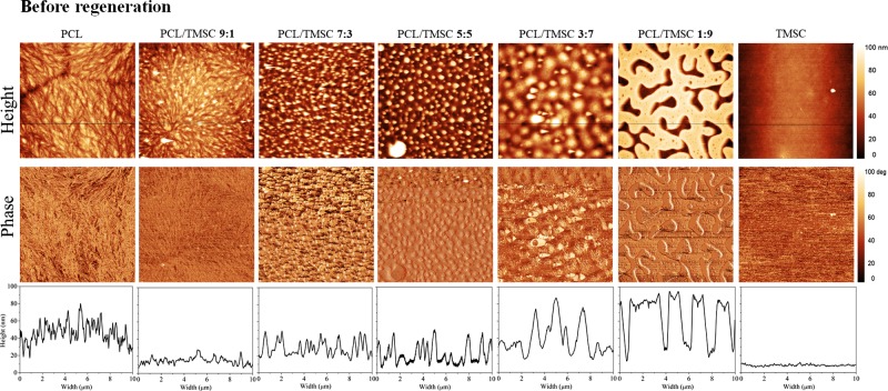
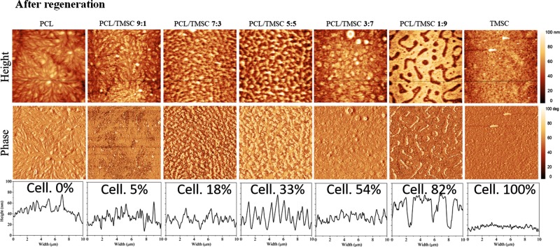
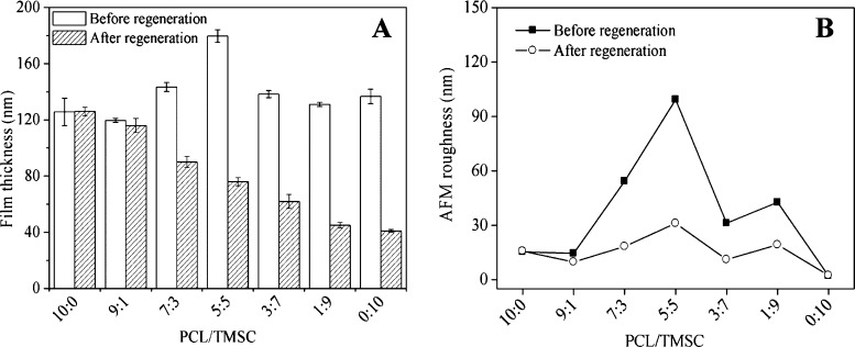
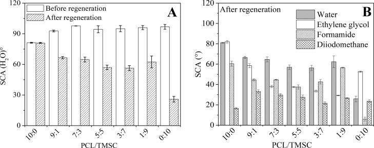
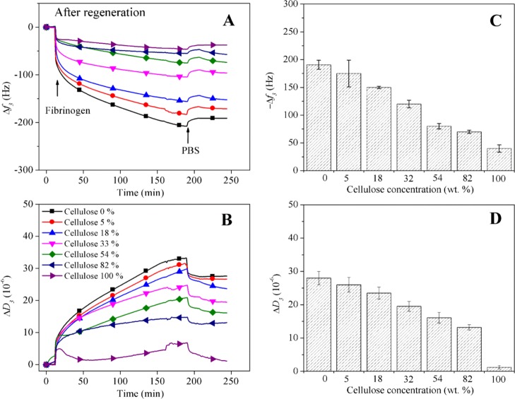
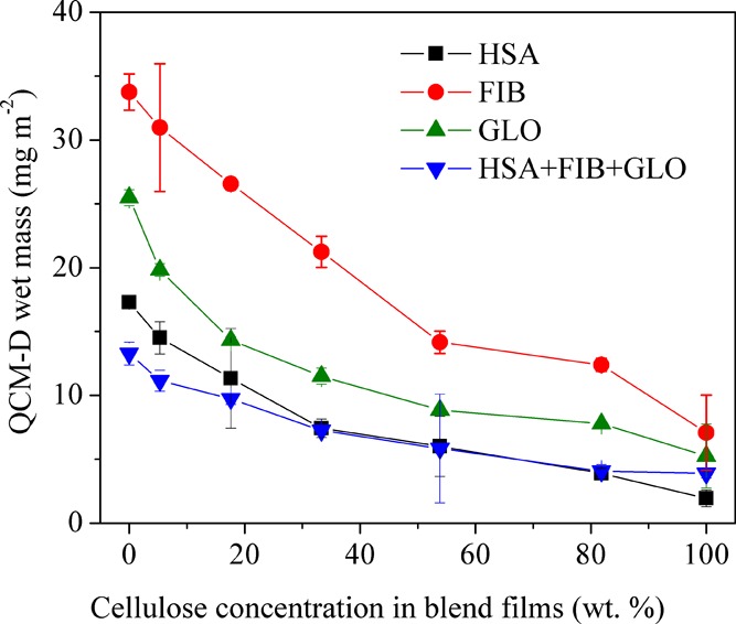
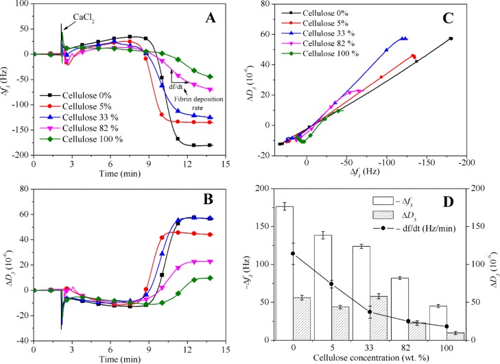
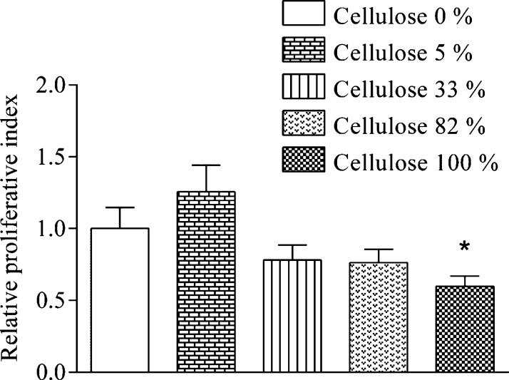
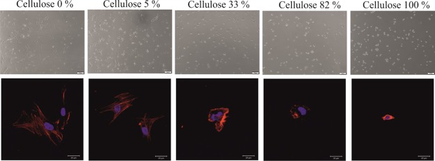
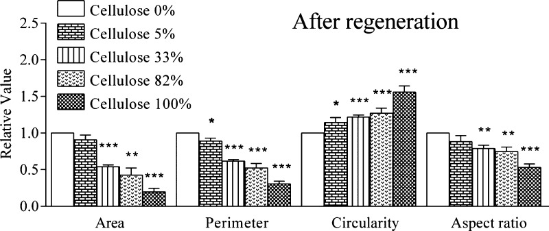
This work describes the interaction of the human blood plasma proteins albumin, fibrinogen, and γ-globulins with micro- and nanopatterned polymer interfaces. Protein adsorption studies were correlated with the fibrin clotting time of human blood plasma and with the growth of primary human pulmonary artery endothelial cells (hECs) on these patterns. It was observed that blends of polycaprolactone (PCL) and trimethylsilyl-protected cellulose form various thin-film patterns during spin coating, depending on the mass ratio of the polymers in the spinning solutions. Vapor-phase acid-catalyzed deprotection preserves these patterns but yields interfaces that are composed of hydrophilic cellulose domains enclosed by hydrophobic PCL. The blood plasma proteins are repelled by the cellulose domains, allowing for a suggested selective protein deposition on the PCL domains. An inverse proportional correlation is observed between the amount of cellulose present in the films and the mass of irreversibly adsorbed proteins. This results in significantly increased fibrin clotting times and lower masses of deposited clots on cellulose-containing films as revealed by quartz crystal microbalance with dissipation measurements. Cell viability of hECs grown on these surfaces was directly correlated with higher protein adsorption and faster clot formation. The results show that presented patterned polymer composite surfaces allow for a controllable blood plasma protein coagulation and a significant biological response from hECs. It is proposed that this knowledge can be utilized in regenerative medicine, cell cultures, and artificial vascular grafts by a careful choice of polymers and patterns.


Conflict of interest statement

The authors declare no competing financial interest.

Figures

Scheme 1
Scheme 1. Nano- and Micropatterned Surfaces from PCL and Cellulose and Their Interaction with Human Blood Plasma Proteins and Primary Human Pulmonary Artery Endothelial Cells
Figure 1
Figure 1
AFM images and cross-sectional profiles of nano- and micropatterned surfaces of PCL/TMSC.
Figure 2
Figure 2
AFM images and cross-sectional profiles of nano- and micropatterned surfaces of PCL/cellulose, obtained from PCL/TMSC by regeneration with HCl vapors. Final cellulose concentrations are shown in wt %.
Figure 3
Figure 3
Profilometry thickness (A) and AFM roughness (B) of nano- and micropattered PCL/TMSC surfaces, before and after regeneration.
Figure 4
Figure 4
Wettability of water (A) and various solvents (B) on nano- and micropattered PCL/TMSC surfaces before and after regeneration.
Figure 5
Figure 5
QCM-D frequency (A) and dissipation (B) changes during the adsorption of FIB on blend films containing increasing amounts of cellulose. The final changes in frequency f3 (C) and dissipation D3 (D) after rinsing with buffer are shown.
Figure 6
Figure 6
Protein adsorption vs cellulose concentration in blend films after regeneration.
Figure 7
Figure 7
Time-dependent change in frequency f3 (A) and dissipation D3, (B) during blood plasma coagulation on films containing increasing amounts of cellulose. (C) f3 vs D3 for the same experiments. (D) Negative f3 and positive D3 shifts 14 min after the addition of CaCl2 including the fibrin deposition rate (df/dt) on films with increasing amounts of cellulose.
Figure 8
Figure 8
Viability of hECs cultured on PCL/cellulose surfaces.
Figure 9
Figure 9
Changes in cellular morphology of hECs cultured on films containing increasing amounts of cellulose visualized in bright field (top) and fluorescence microscopy after staining of F-actin (bottom).
Figure 10
Figure 10
Quantification of changes in hECs’ cellular morphology induced by blend films containing increasing amounts of cellulose. For each sample, n ≥ 50 cells were analyzed. Circularity was defined as [4π(cell area)/(cell perimeter)2]. The aspect ratio was defined as the ratio between the long axis of the cell and the longest axis perpendicular to the long axis.

References

    1. Woodruff M. A.; Hutmacher D. W. The return of a forgotten polymer—Polycaprolactone in the 21st century. Prog. Polym. Sci. 2010, 35, 1217–1256. 10.1016/j.progpolymsci.2010.04.002. - DOI
    1. Markstedt K.; Mantas A.; Tournier I.; Martínez Ávila H.; Hägg D.; Gatenholm P. 3D Bioprinting Human Chondrocytes with Nanocellulose–Alginate Bioink for Cartilage Tissue Engineering Applications. Biomacromolecules 2015, 16, 1489–1496. 10.1021/acs.biomac.5b00188. - DOI - PubMed
    1. Klemm D.; Heublein B.; Fink H.-P.; Bohn A. Cellulose: Fascinating Biopolymer and Sustainable Raw Material. Angew. Chem., Int. Ed. 2005, 44, 3358.10.1002/anie.200460587. - DOI - PubMed
    1. Dugan J. M.; Gough J. E.; Eichhorn S. J. Bacterial cellulose scaffolds and cellulose nanowhiskers for tissue engineering. Nanomedicine 2013, 8, 287–298. 10.2217/nnm.12.211. - DOI - PubMed
    1. Lombardo S.; Thielemans W. Thermodynamics of adsorption on nanocellulose surfaces. Cellulose 2019, 26, 249–279. 10.1007/s10570-018-02239-2. - DOI

Publication types