. 2019 May 9;19(1):91.
doi: 10.1186/s12866-019-1457-z.

Infection and nuclear interaction in mammalian cells by 'Candidatus Berkiella cookevillensis', a novel bacterium isolated from amoebae

Affiliations

Infection and nuclear interaction in mammalian cells by 'Candidatus Berkiella cookevillensis', a novel bacterium isolated from amoebae

Nicholas B Chamberlain et al. BMC Microbiol. .

Abstract

Background: 'Candidatus Berkiella cookevillensis' and 'Ca. Berkiella aquae' have previously been described as intranuclear bacteria of amoebae. Both bacteria were isolated from amoebae and were described as appearing within the nuclei of Acanthamoeba polyphaga and ultimately lysing their host cells within 4 days. Both bacteria are Gammaproteobacteria in the order Legionellales with the greatest similarity to Coxiella burnetii. Neither bacterium grows axenically in artificial culture media. In this study, we further characterized 'Ca. B. cookevillensis' by demonstrating association with nuclei of human phagocytic and nonphagocytic cell lines.

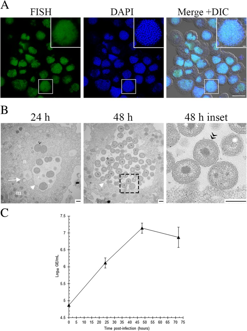
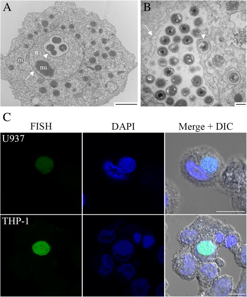
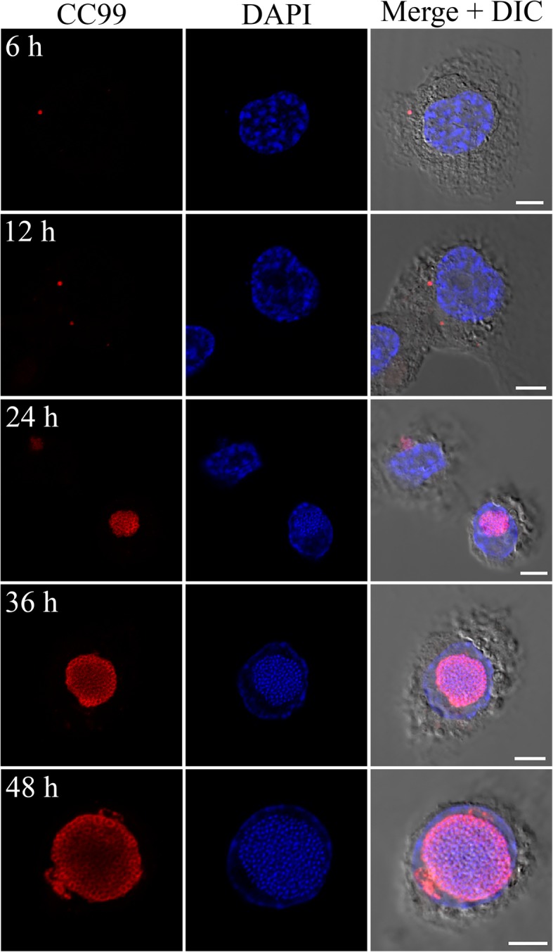
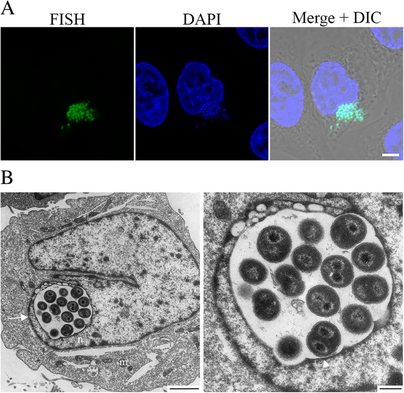
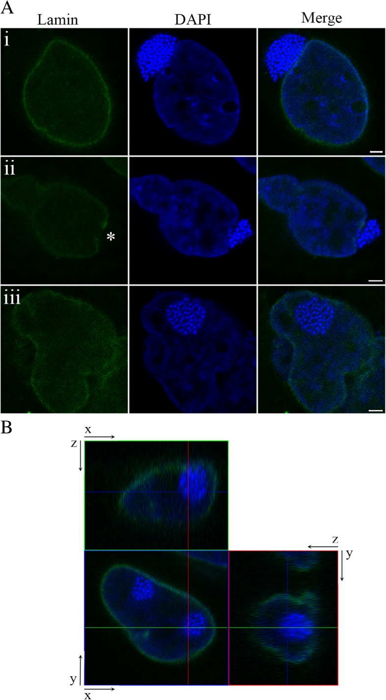
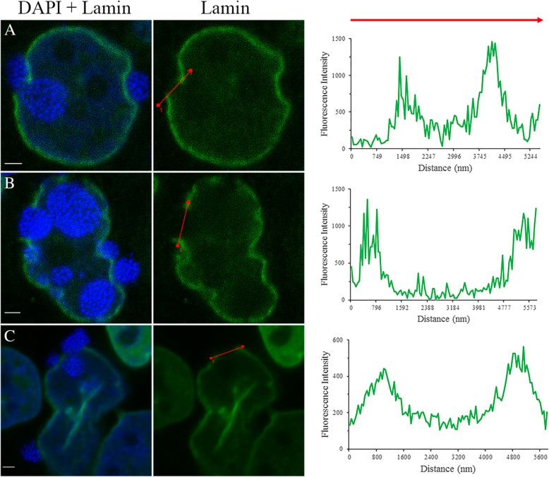
Results: Transmission electron microscopy (TEM) and confocal microscopy were used to confirm nuclear co-localization of 'Ca. B. cookevillensis' in the amoeba host A. polyphaga with 100% of cells having bacteria co-localized with host nuclei by 48 h. TEM and confocal microscopy demonstrated that the bacterium was also observed to be closely associated with nuclei of human U937 and THP-1 differentiated macrophage cell lines and nonphagocytic HeLa human epithelial-like cells. Immunofluorescent staining revealed that the bacteria-containing vacuole invaginates the nuclear membranes and appears to cross from the cytoplasm into the nucleus as an intact vacuole.

Conclusion: Results of this study indicate that a novel coccoid bacterium isolated from amoebae can infect human cell lines by associating with the host cell nuclei, either by crossing the nuclear membranes or by deeply invaginating the nuclear membranes. When associated with the nuclei, the bacteria appear to be bound within a vacuole and replicate to high numbers by 48 h. We believe this is the first report of such a process involving bacteria and human cell lines.

Keywords: Bacteria; Coxiella; Endosymbiont; Human; Legionella; Nucleus; Symbiosis.


Conflict of interest statement

Ethics approval and consent to participate

Not applicable.

Consent for publication

Not applicable.

Competing interests

The authors declare they have no competing interests.

Publisher’s Note

Springer Nature remains neutral with regard to jurisdictional claims in published maps and institutional affiliations.

Figures

Fig. 1
Fig. 1
Growth of ‘Ca. Berkiella cookevillensis’ in Acanthamoeba polyphaga. A. polyphaga was infected for 48 h with ‘Ca. B. cookevillensis’ at an MOI of 1. a Intracellular bacteria were detected using confocal microscopy with helper and 6-FAM-labeled FISH probes specific for the 16S rRNA of the bacterium (green). DAPI staining was used to visualize the amoebal nucleus but also stains the bacteria (blue). Large numbers of bacteria are co-localized with nuclei of amoebae by 48 h p.i. Bars, 10 μm. b TEMs showing the growth of ‘Ca. B. cookevillensis’ following infection for 24 and 48 h in A. polyphaga at an MOI of 1. At 24 h, small numbers of bacteria are visible within the double-membraned (white arrow) nucleus (n). The euchromatin of the nucleoplasm appears more electron-lucent than the cytoplasm. Mitochondria (m) are visible in the cytoplasm. The bacteria are most often seen within an electron-translucent space surrounded by a darkened, single membrane (white triangle). More than one bacteria-containing vacuole often appears within a nucleus. On occasion, no electron-translucent space or membrane is visualized around the bacteria, as for the bacterium at 24 h (indicated by >). By 48 h, increased numbers of bacteria are within bacteria-containing vacuoles and dividing bacteria are evident (*). The gram-negative outer membrane can also be distinguished in TEMs as is indicated in the magnified insets at 48 h (>>). Bars, 500 nm. c Bacterial growth of ‘Ca. B. cookevillensis’ in A. polyphaga as confirmed by qPCR. ‘Ca. B. cookevillensis’ increased by 1.5 log10 when infected at an MOI of 1. Data represent means of two independent experiments performed in triplicate
Fig. 2
Fig. 2
Growth of ‘Ca. B. cookevillensis’ in human macrophage-like cells. TEMs of ‘Ca. B. cookevillensis’ appearing to be within the nucleus (n) of human U937 macrophages at 24 h (a) and 48 h (b) following infection with an MOI of 10. At 24 h, bacteria are contained within the nucleoplasm surrounded by electron-translucent space within a vacuole (white triangle). The bilayer of the nuclear envelope is visible (white arrow). The nucleolus (nu) is also visible. By 48 h, larger numbers of bacteria are visible in the nuclear vacuoles and the vacuole fills the nucleus. A cytoplasmic vacuole with a single bacterium is also visible. c Confocal micrographs of FISH stains using probes specific for ‘Ca. B. cookevillensis’ in U937 cells (top) and an additional human macrophage cell line, THP-1 (bottom) at 48 h p.i. Nuclei and bacteria are also stained with DAPI. Inclusions filled with bacteria are visible for both cell lines. Bars, 10 μm
Fig. 3
Fig. 3
Time course of ‘Ca. B. cookevillensis’ infection in PMA-differentiated THP-1 macrophage-like cells. THP-1 cells were infected with ‘Ca. B. cookevillensis’ (CC99) for 1 h at an MOI of 10 and incubated for the designated amount of time. Following incubation, immunofluorescent staining of bacteria was conducted with rabbit anti-serum to ‘Ca. B. cookevillensis’ and a TRITC-conjugated secondary antibody prior to DAPI staining. At 6 h p.i., puncta of individual or small clusters of bacteria are visible in the cytoplasm. By 12–24 h, the bacterial inclusions appear perinuclear or co-localized with the nucleus. At 36–48 h, the bacteria-containing vacuoles are filled with bacteria, and appear to be enclosed within the nuclear envelope. Imaging was performed with laser scanning confocal microscopy. Bars, 5 μm
Fig. 4
Fig. 4
Growth of ‘Ca. Berkiella cookevillensis’ in non-phagocytic HeLa cells. a Confocal micrographs of FISH staining with 6-FAM-conjugated probe and helper probes targeting the 16S rRNA of ‘Ca. B. cookevillensis’ confirms the infectivity of ‘Ca. B. cookevillensis’ for HeLa human epithelial-like cells. Bar, 5 μm. b TEM images of bacteria appearing to be within the nucleus (n) of a HeLa cell. The left image (bar, 2 μm) shows the location of the nuclear envelope (white arrow). Mitochondria (m) are visible in the cytoplasm. The bacteria appear surrounded by electron-translucent space within a vacuole. The vacuolar membrane (white triangle) is visible in the enlarged image (right) with the vacuole surrounded by loose euchromatin. Dividing cells can also be seen in the image (bar, 500 nm)
Fig. 5
Fig. 5
Time course of ‘Ca. Berkiella cookevillensis’ infection in HeLa cells. HeLa cells were infected with ‘Ca. B. cookevillensis’ for 1 h at an MOI of 200, and then incubated for the designated amount of time. Following the 1 h incubation (time 0), bacterial and mammalian DNA was stained with DAPI. Imaging was performed with laser scanning confocal microscopy. After only 1 h of incubation, distinct bacteria can be found in the cytoplasm. By 3 h p.i., bacteria are still within the cytoplasm, but by 6–12 h, bacteria are more perinuclear. At 24 h, larger numbers of bacteria are present within cytoplasmic vacuoles and nuclear-associated vacuoles. Bars, 5 μm
Fig. 6
Fig. 6
Lamin staining of ‘Ca. Berkiella cookevillensis’-treated HeLa cells. Indirect immunofluorescent staining of lamin A/C (green) performed in conjunction with a DAPI stain (blue) for HeLa cell and bacterial DNA. a Representative confocal micrographs of HeLa cells infected with ‘Ca. B. cookevillensis’ at an MOI of 200 for 24 h demonstrate the invasion process of bacteria-containing vacuoles into the host nucleus (i-iii). (i) Image depicts the bacteria-containing vacuole localized to the juxtanuclear region of the HeLa cell with lamin staining indicating an intact nuclear lamina. (ii) Entry of the ‘Ca. B. cookevillensis’ vacuole into the nucleus. Asterisk (*) denotes site of nucleus invasion with a disruption of lamin staining. (iii) Intact bacteria-containing vacuole within the nucleus of the host cell with no indication of lamin staining of the ‘Ca. B. cookevillensis’ vacuole. Bars, 2 μm. b z-y and z-x projections demonstrate the bacteria-containing vacuole within the nucleus
Fig. 7
Fig. 7
Detection of lamina gaps in nuclei of ‘Ca. Berkiella cookevillensis’-treated HeLa cells. Indirect immunofluorescence staining of HeLa cells infected with ‘Ca. B. cookevillensis (CC99) at an MOI of 200 for 24 h. HeLa cells were labeled using an anti-lamin A/C antibody and co-stained with DAPI. a-c Three examples of CC99-rich vacuoles invading the host cell nucleus. The DAPI + lamin merged micrograph demonstrates the position of bacterial DNA to the nuclear lamina. The fluorescence intensity profile for the lamin stain was constructed to quantify the size of the nuclear lamina gap (red arrow). Bars, 2 μm
Fig. 8
Fig. 8
Exclusion of cytoplasmic markers during ‘Ca. Berkiella cookevillensis’ nuclear invasion of HeLa cells. HeLa cells were transduced to express Rab5a-GFP (green) prior to CC99 infection. In these images Rab5a-GFP not associated with early endosomes was used as a cytoplasm marker. Cells were infected at an MOI of 200 and incubated for 24 h. HeLa cell and bacterial DNA were stained with DAPI (blue). Image series demonstrates a CC99-containing vacuole in close proximity to the host nucleus, b invading the host nucleus, and c inside the host nucleus. Throughout the nuclear invasion process, the cytoplasmic marker Rab5a-GFP was excluded from the nucleus, nor was there any DAPI-stained chromatin in the cytoplasm. Bars, 2 μm

References

    1. Mehari YT, Hayes BJ, Redding KS, Mariappan PV, Gunderson JH, Farone AL, Farone MB. Description of ‘Candidatus Berkiella aquae’ and ‘Candidatus Berkiella cookevillensis’, two intranuclear bacteria of freshwater amoebae. Int J Syst Evol Microbiol. 2016;66:536–541. doi: 10.1099/ijsem.0.000750. - DOI - PubMed
    1. Mehari YT, Arivett BA, Farone AL, Gunderson JH, Farone MB. Draft genome sequences of two novel amoeba-resistant intranuclear bacteria, “Candidatus Berkiella cookevillensis” and “Candidatus Berkiella aquae”. Genome Announc. 2016;4:e01732–e01715. doi: 10.1128/genomeA.01732-15. - DOI - PMC - PubMed
    1. Schulz F, Horn M. Intranuclear bacteria: inside the cellular control center of eukaryotes. Trends Cell Biol. 2015;25:339–346. doi: 10.1016/j.tcb.2015.01.002. - DOI - PubMed
    1. Kusch J, Görtz H-D. Towards an understanding of the killer trait: Caedibacter endosymbionts in Paramecium. Prog Mol Subcell Biol. 2006;41:61–76. doi: 10.1007/3-540-28221-1_4. - DOI - PubMed
    1. Schulz F, Lagkouvardos I, Wascher F, Aistlietner K, Kostanjšek R, Horn M. Life in an unusual niche: a bacterial symbiont infecting the nucleus of amoebae. ISME J. 2014;8:1634–1644. doi: 10.1038/ismej.2014.5. - DOI - PMC - PubMed

Publication types

LinkOut - more resources