. 2019 May 9;19(1):89.
doi: 10.1186/s12890-019-0857-z.

Robotic assisted-bronchoscopy: technical tips and lessons learned from the initial experience with sampling peripheral lung lesions

Affiliations

Robotic assisted-bronchoscopy: technical tips and lessons learned from the initial experience with sampling peripheral lung lesions

Septimiu Dan Murgu. BMC Pulm Med. .

Abstract

Background: Peripheral pulmonary nodules are increasingly detected in patients screened for lung cancer or during disease progression of thoracic or extrathoracic malignancies. Sampling these lesions requires surgery, computed tomography (CT)-guided biopsy or bronchoscopic interventions. Bronchoscopic interventions are preferable because they have lower complications and often patients may not be ideal candidates for surgical or CT-guided biopsy. In addition, guidelines recommend diagnosis and staging in one single procedure. The diagnostic yield of existing advanced bronchoscopic techniques including electromagnetic navigation, radial probe ultrasonography, ultrathin bronchoscopy or virtual bronchoscopy remains suboptimal. The purpose of this paper is to codify the technique whereby a diagnostic bronchoscopy is performed using the new robotic platform.

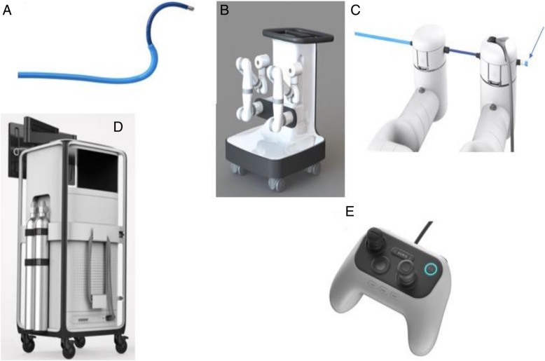
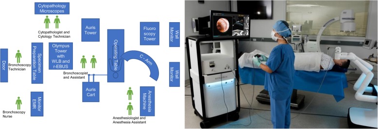
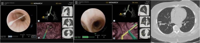
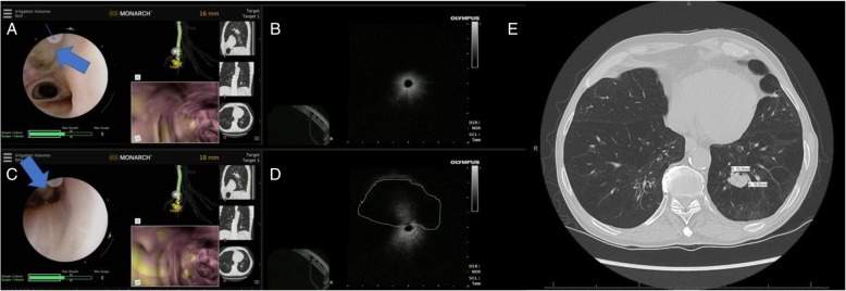
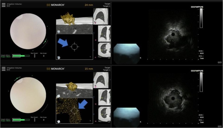
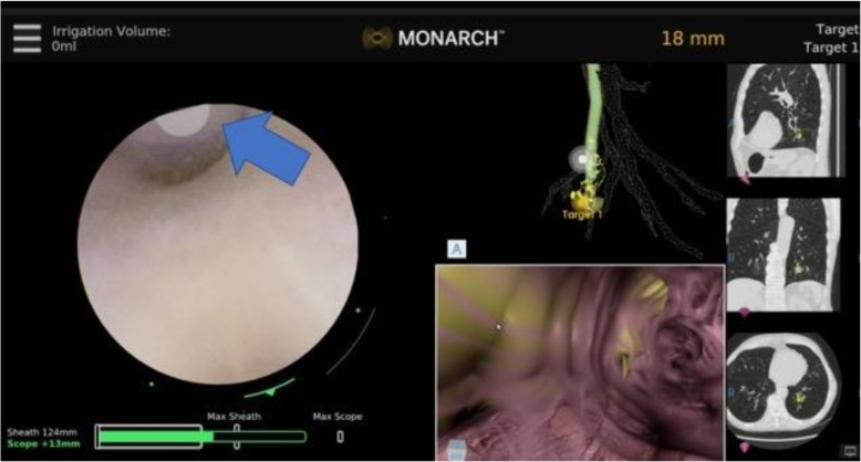
Methods: In the present report, I describe the technique for performing robotic-assisted bronchoscopy (RAB) using the Monarch™ platform (Auris Health, Inc., Redwood City, CA).

Results: Appropriate team training, patient selection, anesthesia settings, optimal tissue acquisition and processing, and prevention of complications are described and illustrated.

Conclusions: RAB may be beneficial for patients with peripheral lung lesions that require biopsy prior to surgical resection, stereotactic radiation, targeted or immunotherapy.

Keywords: Guided bronchoscopy; Lung cancer; Navigation bronchoscopy; Peripheral lung lesion; Peripheral lung nodule; Radial probe ultrasound; Robotic bronchoscopy; Screening.


Conflict of interest statement

Ethics approval and consent to participate

Not applicable.

Consent for publication

Not applicable- no personal information is provided in your manuscript.

Competing interests

The author declares that he has no competing interests.

Publisher’s Note

Springer Nature remains neutral with regard to jurisdictional claims in published maps and institutional affiliations.

Figures

Fig. 1
Fig. 1
a The Auris Bronchoscope is formed by an inner scope and the outer sheath. The Bronchoscope includes a camera that provides the operative perspective, an integrated light source in the handle and a 2.1 mm inner diameter working channel for the passing of manually controlled tools. b The Auris Cart with the robotic arms. c Attachment of the bronchoscope to the robotic arms with the proximal valve for saline, air or instrument insertion (arrow). d The Tower with the monitor for endoscopic and electromagnetic navigation display. e The Controller. Photos courtesy of Auris Health, Inc., Redwood City, CA
Fig. 2
Fig. 2
Schematic representation of the room set up for robotic assisted bronchoscopy (left panel). Real life picture of the Auris tower, cart, operating table and fluoroscopy system (right panel- photo courtesy of Auris Health, Inc., Redwood City, CA.) REBUS: radial probe ultrasound; EMR: electronic medical records; WLB: white light bronchoscopy
Fig. 3
Fig. 3
Manually (in yellow) and automatically (in green) generated pathways to a peripheral nodule in RB8 (anterior basal segment of the right lower lobe). The two pathways overlap in the large central airways (left panel) but clearly diverge in 5th generation airway (middle panel). The bronchoscopist must decide which pathway to follow first. In our practice, we follow the manually generated pathway and confirm it with REBUS once at target. If the nodule is not seen on REBUS, the scope is pulled back and the automatically generated pathway is then followed. The right panel shows the nodule biopsied in this case (a 15.8 X 15.7 mm nodule in RB8) REBUS: radial probe ultrasound
Fig. 4
Fig. 4
REBUS- nodule relationship during robotic bronchoscopy. a REBUS probe (thick arrow) is advanced under direct visualization and seen exiting the scope at the site of the indicator (at 11 o’clock position on the screen- thin arrow). b in that airway, the REBUS image only showed air artifact. c In the same location, the REBUS probe is then oriented towards 6 O’clock position on the screen. The probe is in partial contact with the airway wall (thick arrow). d In that position, REBUS screen shows the nodule as an isoechoic image- highlighted with a dashed line). It is also obvious that nodule as seen on REBUS, although it appears to be at 12 o’clock position relative to the probe, it is clearly at 6’o’clock position as that is where the probe touches the airway wall. This relationship is crucial to identify as the needle orientation is dependent on it. e The panel shows the chest CT scan with the nodule biopsied in this case (a 15.9 X 18.8 mm nodule in RB10) REBUS: radial probe ultrasound
Fig. 5
Fig. 5
Eccentric and concentric REBUS image patterns during robotic bronchoscopy. Top panel: at 24 mm away from the nodule, the REBUS probe was advanced and only an eccentric pattern was obtained; note that the target is not seen on the Monarch EMN display monitor (blue arrow). Bottom panel: the scope is advanced to 15 mm away from the lesion, and the REBUS monitor now shows a concentric pattern (associated with higher diagnostic yield). Note that the nodule is now also seen on the Monarch EMN display monitor (blue arrow). REBUS: radial probe ultrasound; EMN: electromagnetic navigation
Fig. 6
Fig. 6
Needle aspiration during RAB. The Auris needle is seen exiting the bronchoscope at 11 o’clock position (thick arrow). The needle can be re-oriented as necessary, while the bronchoscope is locked in position, but the tip is free to rotate in the desired location. RAB: robotic-assisted bronchoscopy

References

    1. National Lung Screening Trial Research Team. Aberle DR, Adams AM, Berg CD, Black WC, Clapp JD, et al. Reduced lung-cancer mortality with low-dose computed tomographic screening. N Engl J Med. 2011;365:395–409. doi: 10.1056/NEJMoa1102873. - DOI - PMC - PubMed
    1. Yousaf-Khan U, van der Aalst C, de Jong PA, Heuvelmans M, Scholten E, Lammers JW, et al. Final screening round of the NELSON lung cancer screening trial: the effect of a 2.5-year screening interval. Thorax. 2017;72:48–56. doi: 10.1136/thoraxjnl-2016-208655. - DOI - PubMed
    1. Gould MK, Donington J, Lynch WR, Mazzone PJ, Midthun DE, Naidich DP, et al. Evaluation of individuals with pulmonary nodules: when is it lung cancer? Diagnosis and management of lung cancer, 3rd ed: American College of Chest Physicians evidence-based clinical practice guidelines. Chest. 2013;143:e93S–e120S. doi: 10.1378/chest.12-2351. - DOI - PMC - PubMed
    1. Ettinger DS, Aisner DL, Wood DE, Akerley W, Bauman J, Chang JY, et al. NCCN guidelines insights: non-small cell lung Cancer, version 5.2018. J Natl Compr Cancer Netw. 2018;16:807–821. doi: 10.6004/jnccn.2018.0062. - DOI - PubMed
    1. Silvestri GA, Gonzalez AV, Jantz MA, Margolis ML, Gould MK, Tanoue LT, et al. Methods for staging non-small cell lung cancer: diagnosis and management of lung cancer, 3rd ed: American College of Chest Physicians evidence-based clinical practice guidelines. Chest. 2013;143:e211S–e250S. doi: 10.1378/chest.12-2355. - DOI - PubMed