. 2019 May 9;18(1):45.
doi: 10.1186/s12940-019-0486-2.

Assessment of total, ligand-induced peroxisome proliferator activated receptor γ ligand activity in serum

Affiliations

Assessment of total, ligand-induced peroxisome proliferator activated receptor γ ligand activity in serum

Lariah Edwards et al. Environ Health. .

Abstract

Background: Humans are exposed to a complex mixture of environmental chemicals that impact bone and metabolic health, and traditional exposure assessments struggle to capture these exposure scenarios. Peroxisome proliferator activated receptor-gamma (PPARγ) is an essential regulator of metabolic and bone homeostasis, and its inappropriate activation by environmental chemicals can set the stage for adverse health effects. Here, we present the development of the Serum PPARγ Activity Assay (SPAA), a simple and cost-effective method to measure total ligand activity in small volumes of serum.

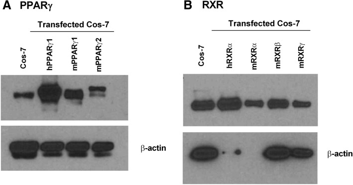
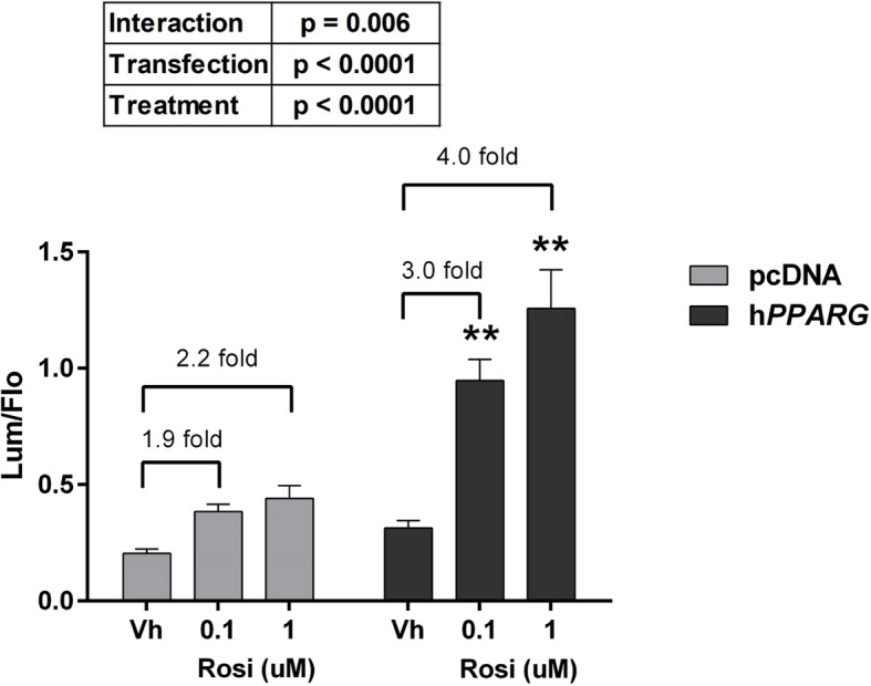
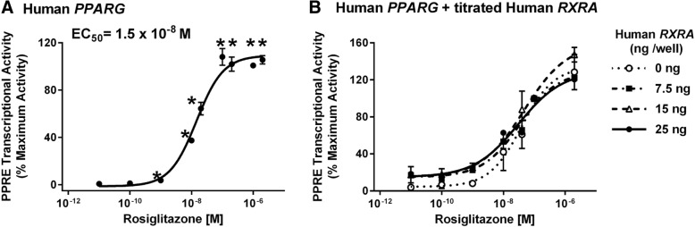
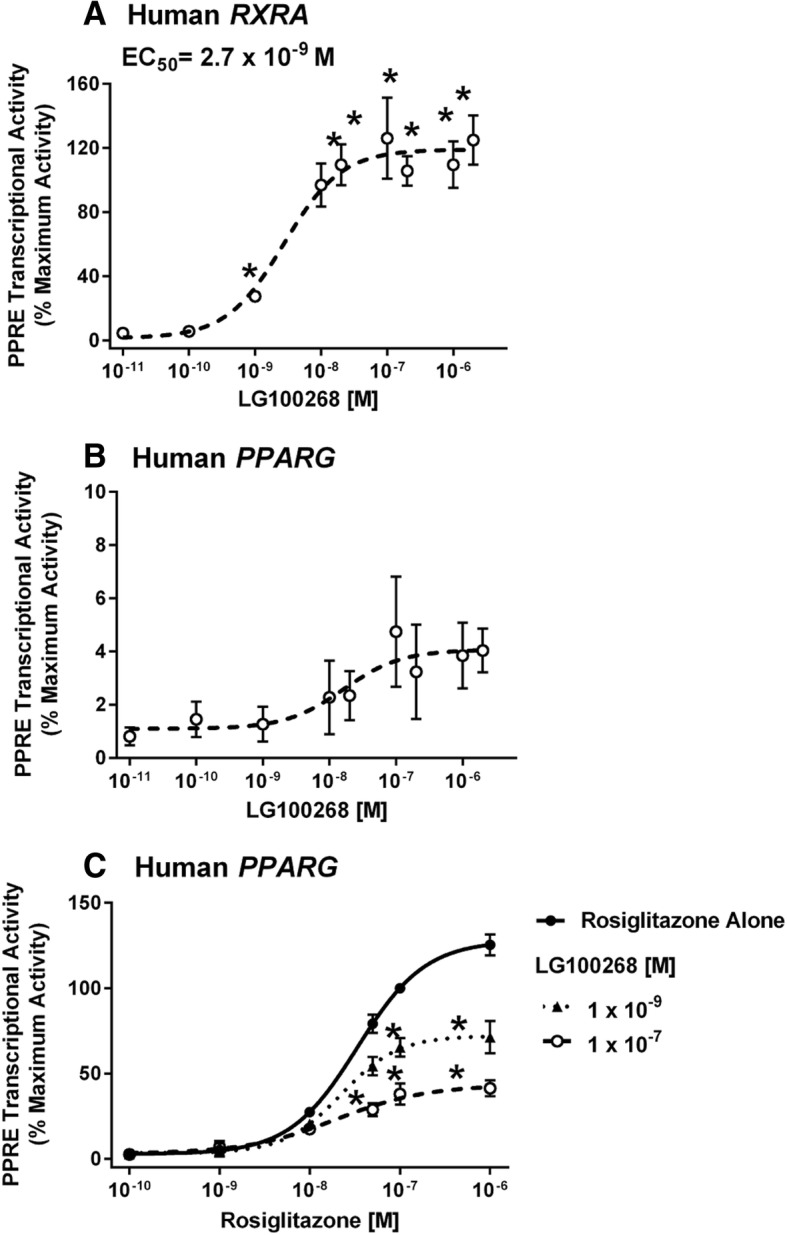
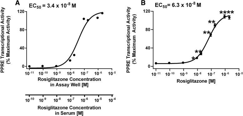
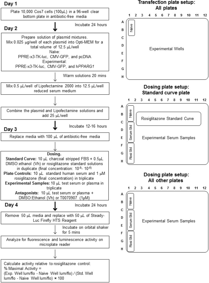
Methods: First, we determined essential components of the bioassay. Cos-7 cells were transfected with combinations of expression vectors for human PPARγ and RXRα, the obligate DNA-binding partner of PPARγ, along with PPRE (DR1)-driven luciferase and control eGFP reporter constructs. Transfected cells were treated with rosiglitazone, a synthetic PPARγ ligand and/or LG100268, a synthetic RXR ligand, to characterize the dose response and determine the simplest and most efficacious format. Following optimization of the bioassay, we assessed the cumulative activation of PPARγ by ligands in serum from mice treated with a PPARγ ligand and commercial human serum samples.

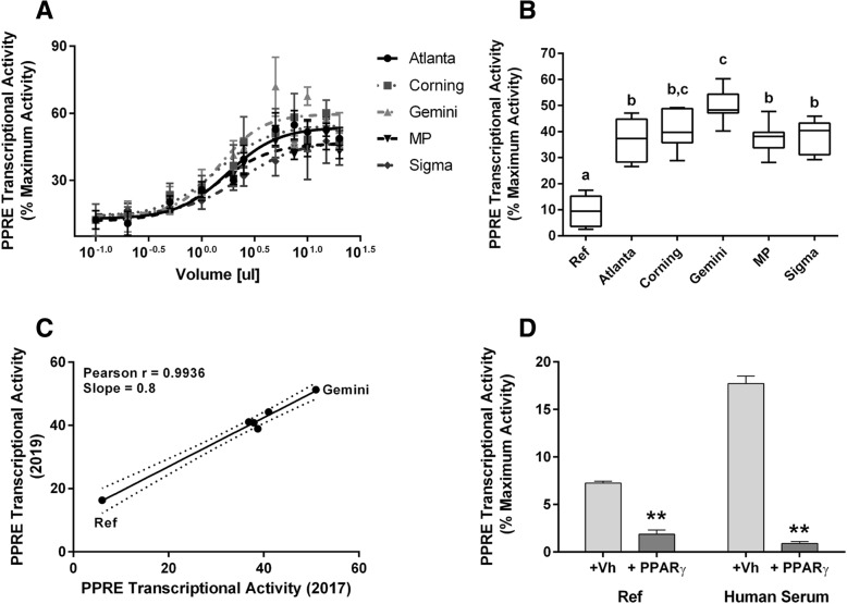
Results: Cos-7 cells endogenously express sufficient RXR to support efficacious activation of transfected PPARγ. Co-transfection of an RXR expression vector with the PPARγ expression vector did not increase PPRE transcriptional activity induced by rosiglitazone. Treatment with an RXR ligand marginally increased PPRE transcriptional activity in the presence of transfected PPARγ, and co-treatment with an RXR ligand reduced rosiglitazone-induced PPRE transcriptional activity. Therefore, the final bioassay protocol consists of transfecting Cos-7 cells with a PPARγ expression vector along with the reporter vectors, applying rosiglitazone standards and/or 10 μL of serum, and measuring luminescence and fluorescence after a 24 h incubation. Sera from mice dosed with rosiglitazone induced PPRE transcriptional activity in the SPAA in a dose-dependent and PPARγ-dependent manner. Additionally, human serum from commercial sources induced a range of PPRE transcriptional activities in a PPARγ-dependent manner, demonstrating the ability of the bioassay to detect potentially low levels of ligands.

Conclusions: The SPAA can reliably measure total PPRE transcriptional activity in small volumes of serum. This system provides a sensitive, straightforward assay that can be reproduced in any cell culture laboratory.

Keywords: Human serum; Metabolism disrupting compounds; Mixtures; PPARγ.


Conflict of interest statement

Ethics approval and consent to participate

Animal studies were reviewed and approved by the Institutional Animal Care and Use Committee at Boston University and performed in an American Association for the Accreditation of Laboratory Animal Care accredited facility (Animal Welfare Assurance Number: A3316–01). All animals were treated humanely and with regard for alleviation of suffering.

Consent for publication

Not applicable.

Competing interests

The authors declare that they have no competing interests.

Publisher’s Note

Springer Nature remains neutral with regard to jurisdictional claims in published maps and institutional affiliations.

Figures

Fig. 1
Fig. 1
Cos-7 cells endogenously express PPARγ1 (A) and RXRs (B), proteins necessary for PPARγ to activate transcription. Whole cell lysates were prepared from Cos-7 cells and Cos-7 cells transfected with (a) mouse Pparg1, mouse Pparg2, human PPARG1 expression vectors, or (b) human RXRA, mouse Rxra, mouse Rxrb, mouse Rxrg expression vectors. Lysates were analyzed for PPARγ, RXRα, and βactin expression by immunoblotting
Fig. 2
Fig. 2
Overexpression of PPARγ significantly increases transcriptional response to rosiglitazone. Cos-7 cells were transfected with reporter plasmids (PPRE-luciferase reporter, CMV-GFP reporter) and empty pcDNA 3.1 or a mouse Pparg2 expression vector. Cells then were treated with Vh (0.5% DMSO) or rosiglitazone (as indicated). Luminescence and fluorescence were measured after 24 h. Data are reported as mean ± standard error (N = 7 independent transfections). Statistical analyses indicated in the box are from a 2-Factor ANOVA. ** Significantly different from Vh (p < 0.01, ANOVA, Dunnett’s)
Fig. 3
Fig. 3
Overexpression of human PPARγ1 alone is sufficient to support robust PPARγ transcriptional activity. Cos-7 cells were transfected with reporter plasmids (PPRE-luciferase reporter, CMV-GFP reporter) and a human PPARG1 expression vector (a) or human PPARG1 and human RXRA expression vectors (b). Cells then were treated with Vh (0.5% DMSO, shown as 10− 11 M) or rosiglitazone (10− 10- 2 × 10− 6 M). Luminescence and fluorescence were measured after 24 h. Data were calculated as described in the Methods. Dose response data are reported as mean ± standard error (N = 3–4 independent transfections). Data were fit with a 3-parameter sigmoid equation. * Significantly different from Vh (p < 0.05, ANOVA, Dunnett’s)
Fig. 4
Fig. 4
RXR ligands do not significantly activate PPARγ, but reduce rosiglitazone-induced, PPRE-dependent transcriptional activity. Cos-7 cells were transfected with reporter plasmids (PPRE-luciferase reporter, CMV-GFP reporter) and a human RXRA expression vector (a) or a human PPARG1 expression vector (b-c). Cells were treated with Vh (0.5% DMSO, reported as 10− 11 M), rosiglitazone (10− 10-2 × 10− 6 M), and/or LG100268 (10− 10- 2 × 10− 6 M). Luminescence and fluorescence were measured after 24 h. Data were calculated as described in the Methods. Data are reported as mean ± standard error (N = 4 independent transfections) * Significantly different from Vh (p < 0.05, ANOVA, Dunnett’s).
Fig. 5
Fig. 5
Standard curve for rosiglitazone in the Serum PPARγ Activity Assay with added serum. Cos-7 cells were transfected with reporter plasmids (PPRE-luciferase reporter, CMV-GFP reporter) and a human PPARG1 expression vector. Charcoal stripped serum (10 μl) was added to each well and then the wells were treated with Vh (0.5% DMSO, shown as 10− 11 M) or rosiglitazone (10− 10 - 2 × 10− 6 M), in duplicate. Luminescence and fluorescence were measured after 24 h. Data were calculated as described in the Methods. Data were fit with a 3-parameter sigmoid equation. (a) Standard curve used to calculate mouse serum rosiglitazone concentrations. The upper X axis indicates the concentrations of rosiglitazone in the total volume in the assay well (10− 10-2 × 10− 6 M). The lower X axis indicates the concentrations of rosiglitazone in the volume of excess serum added to the well (10− 9- 2 × 10− 5 M). (b) Compilation of standard curves performed between 2016 and 2019. Data are reported as mean ± standard error (N = 29 independent transfections). Significantly different from Vh (* p < 0.05, **p < 0.01, ANOVA, Dunnett’s)
Fig. 6
Fig. 6
Serum PPARγ Activity can be detected in whole serum from rosiglitazone-treated mice in a dose-dependent manner. Sera were generated from nine-week-old, female, C57BL/6 J mice were treated by oral gavage with Vh (1% carboxymethylcellulose, 0.1% DMSO) or rosiglitazone (0.1, 1, 10, 100 mg/kg) and euthanized after 1 h. Sera were analyzed in Cos-7 cells transfected with reporter plasmids (PPRE-luciferase reporter, CMV-GFP reporter) and human PPARG1. Experimental wells were treated with 10 μL mouse serum, in duplicate. Luminescence and fluorescence were measured after 24 h. Data were calculated as described in the Methods and were calculated relative to the standard curve shown in Fig. 5. (a) Dose response of serum PPRE transcriptional activity. All mice were treated on the same day. (b) Reproducibility assessment. Mice were treated on three different days, with independently prepared dose solutions. Mice from each experiment are indicated by different symbols. For A and B, individual data are plotted with the mean indicated by a line. Different letters indicate group means that differed significantly, while groups with the same letter did not differ significantly (p < 0.05, ANOVA, Tukey). (c) To test for receptor specificity, wells treated with serum from mice that were exposed to Vh or to 1 mg/kg rosiglitazone were co-treated with Vh (0.5%, 50:50, DMSO:Ethanol), PPARγ antagonist (T0070907, 1 μM) or RXR antagonist (HX 531, 2 μM). For C, data are presented as means ± standard error from all mice in the treatment group. Significantly different from Vh (**p < 0.01, 2-Factor ANOVA, Sidak’s)
Fig. 7
Fig. 7
Determination of optimal serum volume in the SPAA. Cos-7 cells were transfected, and control wells were treated as described in Fig. 5. Experimental wells were treated with 0.5–50 μL of serum. Luminescence and fluorescence were measured after 24 h. Data were calculated as described in the Methods. (a) PPRE transcriptional activity measured in different volumes of serum (Gemini). Different letters indicate group means that differed significantly, while groups with the same letter did not differ significantly (p < 0.05, ANOVA, Tukey). (b) Dose response fit of PPRE transcriptional activity measured in different volumes of serum. Dose response data were fit with a 3-parameter sigmoid equation. Data are reported as mean ± standard error (N = 3 independent transfections)
Fig. 8
Fig. 8
Commercial human serum samples from different sources have distinct PPRE transcriptional activity. Cos-7 cells were transfected with human PPARG1 and treated with Vh or rosiglitazone standards as described in Fig. 5. Experimental wells were treated with 0.2–20 μL human serum. Luminescence and fluorescence were measured after 24 h. Data were calculated as described in the Methods. (a) PPRE transcriptional activity in different volumes of commercial human serum samples. Volume response data were fit with a 3-parameter sigmoid equation. (b) Comparison of PPRE transcriptional activity induced by 10 μL of commercial human serum samples. “Ref” refers wells that received 10 μL of stripped fetal bovine serum. Data are reported as mean ± standard error (N = 7 independent transfections; 4 runs in 2017 and 3 runs in 2019). Different letters indicate group means that differed significantly, while groups with the same letter did not differ significantly (p < 0.05, ANOVA, Tukey). (c) Correlation analysis of assay results from 2017 and 2019. (d) To test for receptor specificity, serum-treated wells were co-treated with Vh (0.5%, 50:50,  DMSO:Ethanol) or PPARγ antagonist (T0070907, 1 μM). Data are presented as means ± standard error from all human serum samples (N = 6) or 4 replicates of Ref serum. Significantly different from Vh (** p < 0.01, 2-Factor ANOVA, Sidak’s)
Fig. 9
Fig. 9
Flowchart and sample plate designs for performing the Serum PPARγ Activity Assay

References

    1. Thayer KA, Heindel JJ, Bucher JR, Gallo MA. Role of environmental chemicals in diabetes and obesity: a national toxicology program workshop review. Environ Health Perspect. 2012;120(6):779–789. - PMC - PubMed
    1. Pool R, Rusch E, Institute of Medicine (U.S.). roundtable on environmental health sciences research and medicine: identifying and reducing environmental health risks of chemicals in our society : workshop summary. Washington, D.C.: the National Academies Press; 2014. - PubMed
    1. Myth versus fact about chemicals in commerce. [http://www.socma.com/advocacy/issues/chemical-risk-management/myth-versu...].
    1. Heindel Jerrold J., Blumberg Bruce, Cave Mathew, Machtinger Ronit, Mantovani Alberto, Mendez Michelle A., Nadal Angel, Palanza Paola, Panzica Giancarlo, Sargis Robert, Vandenberg Laura N., vom Saal Frederick. Metabolism disrupting chemicals and metabolic disorders. Reproductive Toxicology. 2017;68:3–33. - PMC - PubMed
    1. Anghel SI, Bedu E, Vivier CD, Descombes P, Desvergne B, Wahli W. Adipose tissue integrity as a prerequisite for systemic energy balance: a critical role for peroxisome proliferator-activated receptor gamma. J Biol Chem. 2007;282(41):29946–29957. - PubMed

Publication types