Review
. 2019 May 14;73(18):2328-2344.
doi: 10.1016/j.jacc.2019.02.045.

Arrhythmia-Induced Cardiomyopathy: JACC State-of-the-Art Review

Affiliations
Review

Arrhythmia-Induced Cardiomyopathy: JACC State-of-the-Art Review

Jose F Huizar et al. J Am Coll Cardiol. .

Abstract

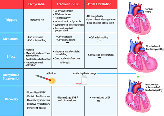
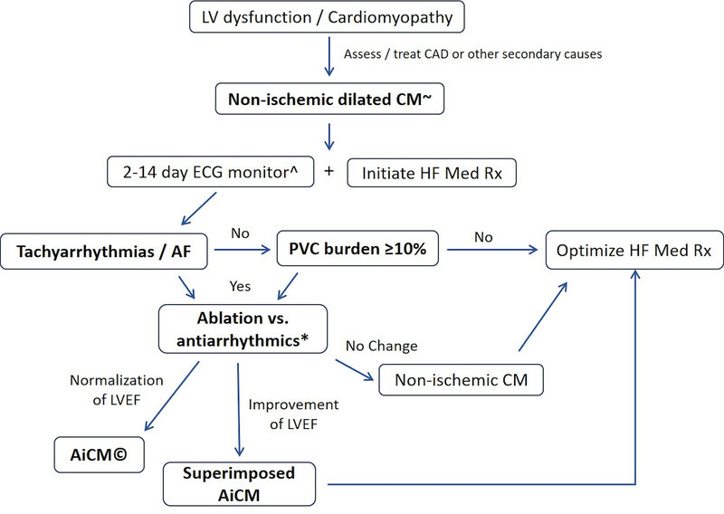
Arrhythmias coexist in patients with heart failure (HF) and left ventricular (LV) dysfunction. Tachycardias, atrial fibrillation, and premature ventricular contractions are known to trigger a reversible dilated cardiomyopathy referred as arrhythmia-induced cardiomyopathy (AiCM). It remains unclear why some patients are more prone to develop AiCM despite similar arrhythmia burdens. The challenge is to determine whether arrhythmias are fully, partially, or at all responsible for an observed LV dysfunction. AiCM should be suspected in patients with mean heart rate >100 beats/min, atrial fibrillation, and/or premature ventricular contractions burden ≥10%. Reversal of cardiomyopathy by elimination of the arrhythmia confirms AiCM. Therapeutic choice depends on the culprit arrhythmia, patient comorbidities, and preferences. Following recovery of LV function, patients require continued follow-up if an abnormal myocardial substrate is present. Appropriate diagnosis and treatment of AiCM is likely to improve quality of life and clinical outcomes and to reduce hospital admission and health care spending.

Keywords: arrhythmia; cardiomyopathy; heart failure; left ventricular dysfunction; premature ventricular contractions; supraventricular tachycardia.


Figures

Central Illustration.
Central Illustration.. Arrhythmia-induced Cardiomyopathies: Possible Triggers, Mediators, Effect, and Recovery.
Figure 1.
Figure 1.
Representative examples of patients with AF with late gadolinium enhancement (LGE) in a patient without (A) and with AF-CM (B). Top panels demonstrate the presence (A) and absence (B) of scar in cardiac MR with late-gadolinium enhancement (LGE) in a patient without and with AF-CM, respectively. Panel C demonstrate change in LVEF from baseline stratified by the presence or absence of scar, while Panel D demonstrates correlation between % of scar (LGE) and change in LVEF from baseline after AF ablation (permission obtained Prabhu S, et al. The CAMERA-MRI Study. J Am Coll Cardiol 2017;70:1949–1961) (23).
Figure 1.
Figure 1.
Representative examples of patients with AF with late gadolinium enhancement (LGE) in a patient without (A) and with AF-CM (B). Top panels demonstrate the presence (A) and absence (B) of scar in cardiac MR with late-gadolinium enhancement (LGE) in a patient without and with AF-CM, respectively. Panel C demonstrate change in LVEF from baseline stratified by the presence or absence of scar, while Panel D demonstrates correlation between % of scar (LGE) and change in LVEF from baseline after AF ablation (permission obtained Prabhu S, et al. The CAMERA-MRI Study. J Am Coll Cardiol 2017;70:1949–1961) (23).
Figure 2.
Figure 2.
Representative cases of patients similar high PVC burden from left coronary cusp without (A) and with (B) associated CM (LVEF 40%). PVC-CM has a wider PVC QRS duration (172ms) when compared to preserved LV function (150ms) (permission obtained Carballeira Pol L, et al. Heart Rhythm 2014; 11:299–306) (75). (C) Representative case of PVC-CM with dispersion of CI of 144ms (cutoff of CI dispersion >99ms best identified patients with and without PVC-CM) (permission obtained Kawamura M, et al. J Cardiovasc Electrophysiol 2014;25:756–62) (39).
Figure 2.
Figure 2.
Representative cases of patients similar high PVC burden from left coronary cusp without (A) and with (B) associated CM (LVEF 40%). PVC-CM has a wider PVC QRS duration (172ms) when compared to preserved LV function (150ms) (permission obtained Carballeira Pol L, et al. Heart Rhythm 2014; 11:299–306) (75). (C) Representative case of PVC-CM with dispersion of CI of 144ms (cutoff of CI dispersion >99ms best identified patients with and without PVC-CM) (permission obtained Kawamura M, et al. J Cardiovasc Electrophysiol 2014;25:756–62) (39).
Figure 3.
Figure 3.
Progression of LV ejection fraction in a large animal PVC model after 4 and 8weeks of a progressive incremental PVC burden starting from 0% (baseline) to 7%, 14%, 24%, 33%, and 50%. (p <0.0001, 1-way ANOVA). Even though PVC-CM is not seen until PVC burden of 33%, a decline in LVEF can be noted at lower PVC burdens (Permission obtained Tan AY, et al. Heart Rhythm 2016;13:755–61) (50).
Figure 4.
Figure 4.. Representative PVC-cardiomyopathy.
A 53-year-old man with 21% PVC burden and LVEF of 40% underwent PVC ablation. Successful ablation was achieved at the mid-septal RVOT just above pulmonary valve with a PVC burden of 1.5% after RFA. (A) 12-lead ECG of representative PVC. (B) Baseline CMR demonstrate absence of scar with late-gadolinium enhancement. LVEF normalized to 55% after 3 months, diagnostic of PVC-CM.
Figure 5.
Figure 5.. Proposed management of potential Arrhythmia-induced CM.
Footnotes: (~) Consider following the algorithm even if CAD is documented or worsening of prior CM is noted (superimposed AiCM). (^) Two-week ambulatory Holter is preferred as increases the diagnosis yield of high PVC burden (≥10%). (*) Consider cardiac MR to assess scar burden and predict response to PVC suppression. Short-term observation is reasonable for PVC-CM as 15% of cases may improve without PVC suppression strategy (49). (©) Continue close surveillance and HF med Rx in those with abnormal LV dimensions and presence of scar (cardiac MRI)

Similar articles

Cited by

References

    1. Bozkurt B, Colvin M, Cook J et al. Current Diagnostic and Treatment Strategies for Specific Dilated Cardiomyopathies: A Scientific Statement From the American Heart Association. Circulation 2016;134:e579–e646. - PubMed
    1. Trulock KM, Narayan SM, Piccini JP. Rhythm control in heart failure patients with atrial fibrillation: contemporary challenges including the role of ablation. J Am Coll Cardiol 2014;64:710–21. - PubMed
    1. Marrouche NF, Brachmann J, Andresen D et al. Catheter Ablation for Atrial Fibrillation with Heart Failure. N Engl J Med 2018;378:417–427. - PubMed
    1. Fazio G, Mongiovi M, Sutera L, Novo G, Novo S, Pipitone S. Segmental dyskinesia in Wolff-Parkinson-White syndrome: a possible cause of dilatative cardiomyopathy. Int J Cardiol 2008;123:e31–4. - PubMed
    1. Gossage AMBHJ. On auricular fibrillation. Quarterly Journal of Medicine 1913;6:435–440.

Publication types

MeSH terms