

Mycobacterial trehalose 6,6'-dimycolate induced vascular occlusion is accompanied by subendothelial inflammation
- PMID: 31072690
- PMCID: PMC6626699
- DOI: 10.1016/j.tube.2019.04.019
Mycobacterial trehalose 6,6'-dimycolate induced vascular occlusion is accompanied by subendothelial inflammation
Abstract
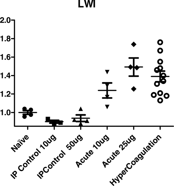
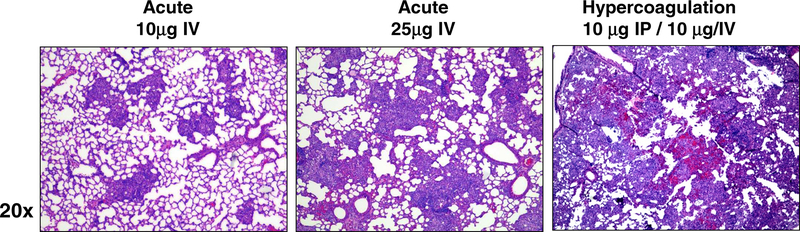
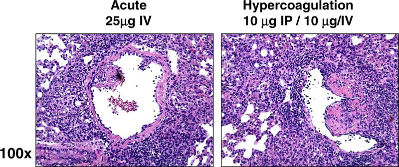
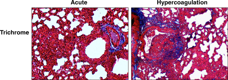
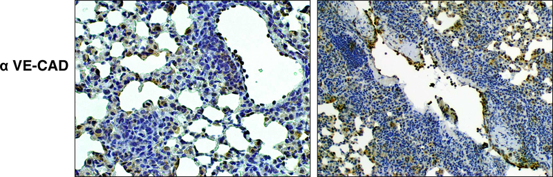
Mycobacterium tuberculosis (MTB) is a pathogen that infects and kills millions yearly. The mycobacterium's cell wall glycolipid trehalose 6,6'-dimycolate (TDM) has been used historically to model MTB induced inflammation and granuloma formation. Alterations to the model can significantly influence the induced pathology. One such method incorporates intraperitoneal pre-exposure, after which the intravenous injection of TDM generates pathological damage effectively mimicking the hypercoagulation, thrombus formation, and tissue remodeling apparent in lungs of infected individuals. The purpose of these experiments is to examine the histological inflammation involved in the TDM mouse model that induces development of the hemorrhagic response. TDM induced lungs of C57BL/6 mice to undergo granulomatous inflammation. Further histological examination of the peak response demonstrated tissue remodeling consistent with hypercoagulation. The observed vascular occlusion indicates that obstruction likely occurs due to subendothelial localized activity leading to restriction of blood vessel lumens. Trichrome staining revealed that associated damage in the hypercoagulation model is consistent with intra endothelial cell accumulation of innate cells, bordered by collagen deposition in the underlying parenchyma. Overall, the hypercoagulation model represents a comparative pathological instrument for understanding mechanisms underlying development of hemorrhage and vascular occlusion seen during MTB infection.
Keywords: Cord Factor; Granuloma; Immunopathology; TDM; Trehalose 6,6′-dimycolate; Tuberculosis.
Copyright © 2019 Elsevier Ltd. All rights reserved.
Figures
Similar articles
-
11beta-hydroxysteroid dehydrogenases are regulated during the pulmonary granulomatous response to the mycobacterial glycolipid trehalose-6,6'-dimycolate.Neuroimmunomodulation. 2009;16(3):147-54. doi: 10.1159/000204227. Epub 2009 Feb 24. Neuroimmunomodulation. 2009. PMID: 19246936
-
Neutrophils Promote Mycobacterial Trehalose Dimycolate-Induced Lung Inflammation via the Mincle Pathway.PLoS Pathog. 2012;8(4):e1002614. doi: 10.1371/journal.ppat.1002614. Epub 2012 Apr 5. PLoS Pathog. 2012. PMID: 22496642 Free PMC article.
-
Trehalose 6,6-Dimycolate from Mycobacterium tuberculosis Induces Hypercoagulation.Am J Pathol. 2016 May;186(5):1221-33. doi: 10.1016/j.ajpath.2015.12.019. Epub 2016 Mar 8. Am J Pathol. 2016. PMID: 26968340
-
Multiple roles of cord factor in the pathogenesis of primary, secondary, and cavitary tuberculosis, including a revised description of the pathology of secondary disease.Ann Clin Lab Sci. 2006 Autumn;36(4):371-86. Ann Clin Lab Sci. 2006. PMID: 17127724 Review.
-
[Pathomorphogenesis of tubercular histologic changes: mechanisms of granuloma formation, maintenance and necrosis].Internist (Berl). 2003 Nov;44(11):1363-73. doi: 10.1007/s00108-003-1036-z. Internist (Berl). 2003. PMID: 14689072 Review. German.
Cited by
-
Recombinant Human Lactoferrin Reduces Inflammation and Increases Fluoroquinolone Penetration to Primary Granulomas During Mycobacterial Infection of C57Bl/6 Mice.Arch Immunol Ther Exp (Warsz). 2022 Feb 28;70(1):9. doi: 10.1007/s00005-022-00648-7. Arch Immunol Ther Exp (Warsz). 2022. PMID: 35226195 Free PMC article.
-
Mycobacterial Trehalose 6,6'-Dimycolate-Induced M1-Type Inflammation.Am J Pathol. 2020 Feb;190(2):286-294. doi: 10.1016/j.ajpath.2019.10.006. Epub 2019 Nov 14. Am J Pathol. 2020. PMID: 31734231 Free PMC article.
References
-
- Sodeinde OA, Subrahmanyam YV, Stark K, Quan T, Bao Y, Goguen JD. A surface protease and the invasive character of plague. Science 1992;258:1004–1007. - PubMed
-
- Robson SC, White NW, Aronson I, Woollgar R, Goodman H, Jacobs P. Acute-phase response and the hypercoagulable state in pulmonary tuberculosis. British journal of haematology 1996;93:943–949. - PubMed
-
- Turken O, Kunter E, Sezer M, Solmazgul E, Cerrahoglu K, Bozkanat E, Ozturk A, Ilvan A. Hemostatic changes in active pulmonary tuberculosis. The international journal of tuberculosis and lung disease : the official journal of the International Union against Tuberculosis and Lung Disease 2002;6:927–932. - PubMed
Publication types
MeSH terms
Substances
Grants and funding
LinkOut - more resources
Full Text Sources
Medical
