. 2019 May 28;116(22):11048-11056.
doi: 10.1073/pnas.1902163116. Epub 2019 May 9.

Elasticity of individual protocadherin 15 molecules implicates tip links as the gating springs for hearing

Affiliations

Elasticity of individual protocadherin 15 molecules implicates tip links as the gating springs for hearing

Tobias F Bartsch et al. Proc Natl Acad Sci U S A. .

Abstract

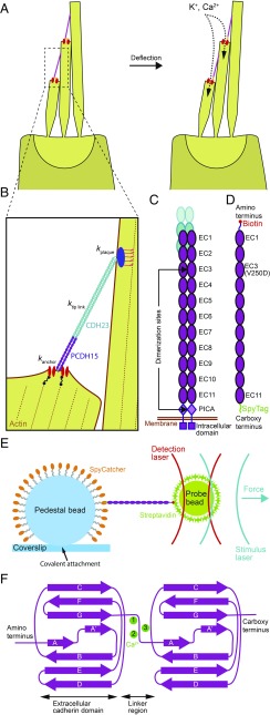
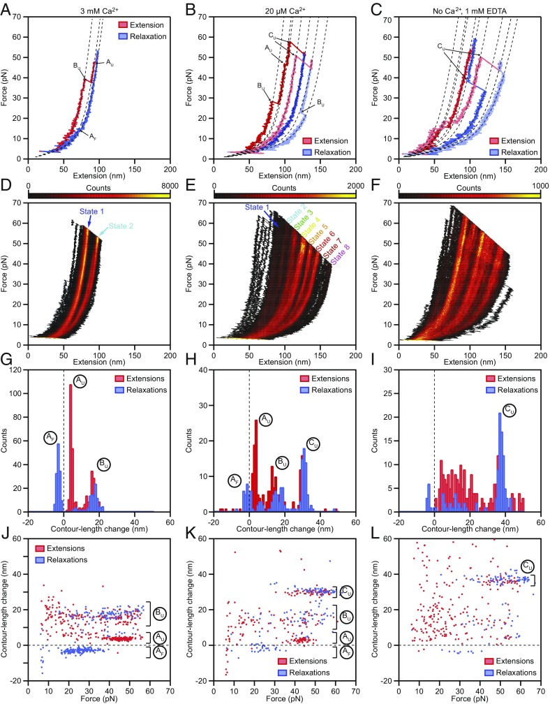
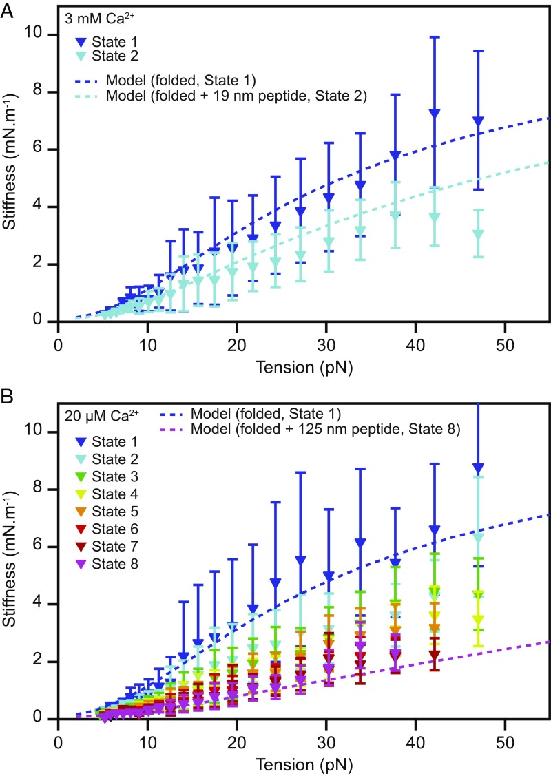
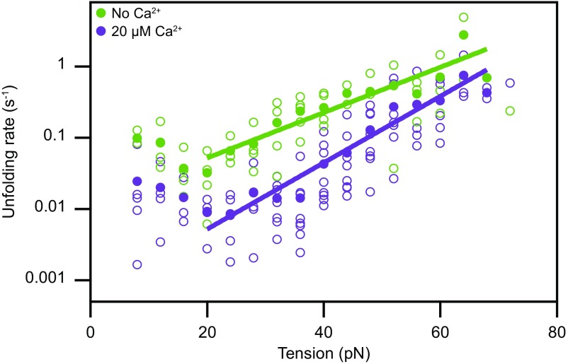
Hair cells, the sensory receptors of the inner ear, respond to mechanical forces originating from sounds and accelerations. An essential feature of each hair cell is an array of filamentous tip links, consisting of the proteins protocadherin 15 (PCDH15) and cadherin 23 (CDH23), whose tension is thought to directly gate the cell's transduction channels. These links are considered far too stiff to represent the gating springs that convert hair bundle displacement into forces capable of opening the channels, and no mechanism has been suggested through which tip-link stiffness could be varied to accommodate hair cells of distinct frequency sensitivity in different receptor organs and animals. Consequently, the gating spring's identity and mechanism of operation remain central questions in sensory neuroscience. Using a high-precision optical trap, we show that an individual monomer of PCDH15 acts as an entropic spring that is much softer than its enthalpic stiffness alone would suggest. This low stiffness implies that the protein is a significant part of the gating spring that controls a hair cell's transduction channels. The tip link's entropic nature then allows for stiffness control through modulation of its tension. We find that a PCDH15 molecule is unstable under tension and exhibits a rich variety of reversible unfolding events that are augmented when the Ca2+ concentration is reduced to physiological levels. Therefore, tip link tension and Ca2+ concentration are likely parameters through which nature tunes a gating spring's mechanical properties.

Keywords: auditory system; entropic stiffness; hair cell; optical trap; vestibular system.


Conflict of interest statement

The authors declare no conflict of interest.

Figures

Fig. 1.
Fig. 1.
The role of tip link proteins in transduction by hair cells. (A) The hair bundle is a cluster of stiff, actin-filled protrusions called stereocilia that stands atop each hair cell in the inner ear. Each stereocilium is connected to its tallest adjacent neighbor through a proteinaceous filament called a tip link (pink), which is coupled at its base to mechanically gated ion channels (red). Deflection of a hair bundle increases the tension in the tip links, biasing the channels toward an open state that allows the influx of positively charged ions. (B) The mechanical element that converts hair bundle displacement into a force capable of opening the channels is called the gating spring. Its stiffness comprises the stiffnesses of the channel and its lower anchor (kanchor), the tip link proteins PCDH15 and CDH23 (ktip link), and the insertional plaque that anchors the link’s top end into the taller stereocilium (kplaque). (C) The mechanical properties of the tip link emerge from its quaternary structure and from the characteristics of its constituent proteins. The lower third of the link consists of a dimer of PCDH15 molecules, each of which includes 11 extracellular cadherin (EC) domains. (D) To measure the mechanical behavior of monomeric PCDH15, we tagged each end with a distinct molecular handle. We eliminated dimerization by a point mutation (V250D) in domain EC3 and by truncation of the PICA domain. (E) We probed the mechanics of a PCDH15 monomer by confining it through molecular handles between an immobile 2-μm glass pedestal bead and a diffusive 1-μm plastic probe bead. To acquire each force-extension relationship, we measured the position of the probe bead with a detection laser while applying a force with a stimulus laser. (F) The folding motifs of individual EC domains influence the mechanical properties of the full-length protein. Up to three calcium ions (green) can bind between successive domains.
Fig. 2.
Fig. 2.
Force-extension measurements of PCDH15 monomers. (A) At a Ca2+ concentration of 3 mM, individual force-extension cycles show two distinct classes of abrupt elongations, the unfolding events AU and BU, as well as refolding events of class AF. The dashed lines represent fits to the trajectories by a protein model. (B) Reducing the Ca2+ concentration to 20 μM elicits an additional class of unfolding events, CU, corresponding to the unfolding of entire cadherin domains. (C) In the absence of Ca2+, unclassifiable structural changes occur in conjunction with the well-defined events CU. (DF) Heatmaps displaying all the force-extension cycles for a single representative molecule at each Ca2+ concentration. The data were binned into pixels of 1 nm × 0.1 pN. A much smaller portion of the state space is accessible at a Ca2+ concentration of 3 mM than at a Ca2+ concentration of 20 µM or in the absence of Ca2+. The heatmaps illustrate which contour lengths of the tethered molecules occurred with increased likelihood during our force-loading protocol as an average over both extensions and relaxations. Prominent regions of elevated occupancy are labeled states 1–8, in which state 1 corresponds to the fully folded protein and state 8 results from the unfolding of three cadherin domains in series with one event of type Bu. (GI) Histograms of the contour length changes of all abrupt elongations verify that these rips can be grouped into classes AF, AU, BU, and CU at Ca2+ concentrations of 3 mM and 20 µM. In the absence of Ca2+, most of the contour length changes are more broadly distributed. (JL) Plots of the contour length change of every rip against the force at which that event occurred revealing the force distributions of each class of structural change. Note that the extensions never completely unfolded a PCDH15 molecule, so elongations could occur even during the relaxation phases. Because the contour lengths observed for the folded protein correspond to the length of monomeric PCDH15 in series with its molecular anchors, the measured extensions exceed those expected for the protein alone. Our analysis corrects for this influence, resulting in values in excellent agreement with the known structure of the protein (Table 1). All force-extension cycles were sampled at intervals of 10 μs and smoothed to a temporal resolution of 1 ms. The waiting times between cycles were 0.2 s for a Ca2+ concentration of 3 mM, 2 s for a Ca2+ concentration of 20 µM, and 4 s in the absence of Ca2+. The number of cycles recorded was 500 for a Ca2+ concentration of 3 mM and 200 for a Ca2+ concentration of 20 µM and in the absence of Ca2+.
Fig. 3.
Fig. 3.
Stiffness of monomeric PCDH15. (A) The stiffnesses of the different conformational states of PCDH15 at a Ca2+ concentration of 3 mM correspond to the slopes of the highly occupied regions of the state space in Fig. 2 D and E and are corrected for the stiffness of the molecular tags and anchors. The dark-blue dashed line represents the stiffness of our model of state 1, the fully folded protein, with the parameter values of Table 1 (b = 3.0 nm; lclinker = 1.35 nm; kfolded = 10 mN·m−1). Parameter values were averaged over both Ca2+ concentrations. The light-blue dashed line represents the model for state 2, with an additional 19-nm segment of unfolded protein with a persistence length of 0.49 nm representing the combined effect of the events AU and BU. (B) The corresponding data for a Ca2+ concentration of 20 μM capture a variety of unfolding events leading to states 2–8. The dark-blue dashed line represents a model of the fully folded protein (state 1); the pink dashed line depicts the modeled stiffness of the protein in state 8, with an unstructured 125-nm-long peptide to represent the unfolding of three cadherin domains in series with contour length changes of 15 nm and 4 nm. The experimental data are mean ± SEM for five molecules at a Ca2+ concentration of 3 mM and for six molecules at a Ca2+ concentration of 20 µM.
Fig. 4.
Fig. 4.
Tension-dependent unfolding rates of a single cadherin domain. Assuming that all domains of PCDH15 are similar and unfold independently, we estimate the rate at which individual EC domains unfold as a function of tension. Domains unfold much more readily in the absence of Ca2+ (green) than at a physiological Ca2+ concentration of 20 µM (purple). The filled circles represent the means for eight molecules at a Ca2+ concentration of 20 µM and for five molecules in the absence of Ca2+. The outlined circles, which represent the data for individual molecules, provide an estimate of the data’s spread. The solid lines are fits of Bells’ model (48) to the data. For 20 µM Ca2+, the unfolding rate with no force is k0 = 0.0006 ± 0.0002 s−1, and the transition-state distance is x = 0.44 ± 0.04 nm; in the absence of Ca2+, these values are k0 = 0.012 ± 0.006 s−1 and x = 0.30 ± 0.04 nm.
Fig. 5.
Fig. 5.
Effect of a hearing loss-associated mutation on PCDH15 mechanics. (A) We deleted V767 in the seventh EC domain of PCDH15. As indicated in the crystal structure (Protein Data Bank ID code 5W1D; image generated with UCSF Chimera), V767 is located in the F strand of the cadherin fold. (B) A state-space heatmap for 500 extension-relaxation cycles reveals that at a Ca2+ concentration of 3 mM the mutant protein can assume four distinct conformational states. The two additional states not observed in the wild-type protein result from unfolding of the pathological cadherin domain in series with the usual states 1 and 2. Unfolding of the pathological domain is rare and occurs in only a few cycles, two of which are superimposed on the heat map (green traces). The waiting time between cycles was 0.2 s.

References

    1. Wiggins P, Phillips R (2004) Analytic models for mechanotransduction: Gating a mechanosensitive channel. Proc Natl Acad Sci USA 101:4071–4076. - PMC - PubMed
    1. Saotome K, et al. (2018) Structure of the mechanically activated ion channel Piezo1. Nature 554:481–486. - PMC - PubMed
    1. Zhao Q, et al. (2018) Structure and mechanogating mechanism of the Piezo1 channel. Nature 554:487–492. - PubMed
    1. Jin P, et al. (2017) Electron cryo-microscopy structure of the mechanotransduction channel NOMPC. Nature 547:118–122. - PMC - PubMed
    1. Corey DP, Hudspeth AJ (1983) Kinetics of the receptor current in bullfrog saccular hair cells. J Neurosci 3:962–976. - PMC - PubMed

Publication types

LinkOut - more resources