. 2019 Mar 28;3(5):1062-1078.
doi: 10.1210/js.2019-00029. eCollection 2019 May 1.

The Catalytic Subunit β of PKA Affects Energy Balance and Catecholaminergic Activity

Affiliations

The Catalytic Subunit β of PKA Affects Energy Balance and Catecholaminergic Activity

Edra London et al. J Endocr Soc. .

Abstract

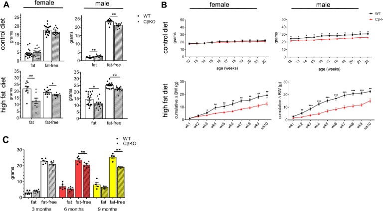
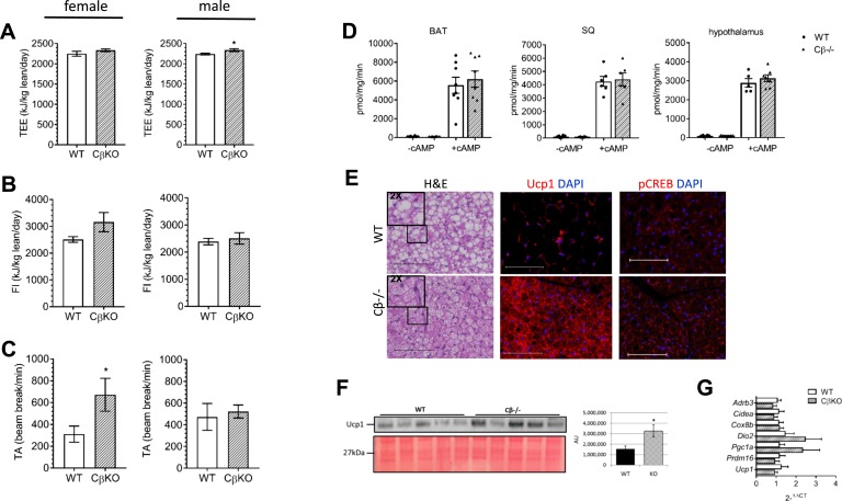
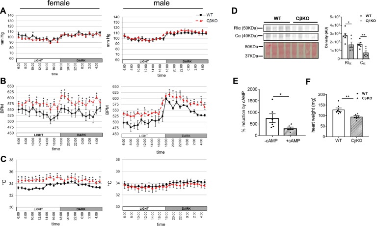
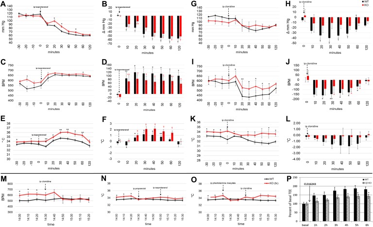
The protein kinase A (PKA) signaling system mediates the effects of numerous hormones, neurotransmitters, and other molecules to regulate metabolism, cardiac function, and more. PKA defects may lead to diverse phenotypes that largely depend on the unique expression profile of the affected subunit. Deletion of the Prkarcb gene, which codes for PKA catalytic subunit β (Cβ), protects against diet-induced obesity (DIO), yet the mechanism for this phenotype remains unclear. We hypothesized that metabolic rate would be increased in Cβ knockout (KO) mice, which could explain DIO resistance. Male, but not female, CβKO mice had increased energy expenditure, and female but not male CβKO mice had increased subcutaneous temperature and increased locomotor activity compared with wild-type (WT) littermates. Urinary norepinephrine (NE) and normetanephrine were elevated in female CβKO mice. CβKO mice had increased heart rate (HR); blocking central NE release normalized HR to that of untreated WT mice. Basal and stimulated PKA enzymatic activities were unchanged in adipose tissue and heart and varied in different brain regions, suggesting that Prkacb deletion may mediate signaling changes in specific brain nuclei and may be less important in the peripheral regulation of PKA expression and activity. This is a demonstration of a distinct effect of the PKA Cβ catalytic subunit on catecholamines and sympathetic nerve signaling. The data provide an unexpected explanation for the metabolic phenotype of CβKO mice. Finally, the sexual dimorphism is consistent with mouse models of other PKA subunits and adds to the importance of these findings regarding the PKA system in human metabolism.

Keywords: PKA; cardiovascular; catecholamines; energy balance; sympathetic outflow.


Figures

Figure 1.
Figure 1.
Prkacb mutants had altered weight gain and body composition as a result of chronic HFD and CD feeding that was sex dependent. (A) Fat mass and fat-free mass of female and male CβKO mice were measured by Echo-MRI after chronic feeding with either CD or HFD (n = 6 to 21/group). (B) Growth curves of female and male WT and CβKO mice during 10-wk CD or HFD feeding (n = 8 to 14/group/sex); body weight data for the HFD feeding study are shown as cumulative change in body weight from baseline. (C) Body composition of male WT and CβKO littermates at 3, 6, and 9 mo of age (n = 6 to 8/group); all data represent means ± SEM. *P < 0.05; **P < 0.01; ***P < 0.001. For statistical analysis, (A) two-tailed paired Student t tests and (B and C) repeated-measures two-way ANOVA with Tukey post hoc test and multiple paired t tests were performed.
Figure 2.
Figure 2.
Metabolic rate and energy in CβKO mice compared with WT littermates. (A) TEE, (B), food intake (FI), and (C) TA during CD feeding were measured in individually housed female and male mice for 4 d at ambient (22°C) temperature; values were adjusted for lean body mass (in kilograms; n = 5 to 7/group/sex). (D) Representative PKA enzymatic activity in BAT, SQ AT (inguinal WAT), and hypothalamus, with and without cAMP stimulation (5 μM; n = 6/group, females). (E) Representative hematoxylin and eosin (H&E) and immunofluorescent staining of BAT for Ucp1 and pCREB (Ser133). Original scale bars represent 100 μM (n = 3/group, males). Inset H&E images are 2X magnified from the areas indicated. (F) Representative Western blot for Ucp1 and densitometry analysis in BAT (n = 5/group, females). (G) Relative mRNA expression of several important regulatory BAT genes was quantified by qRT-PCR. Fold change calculated as 2ΔΔCT. Actb was the housekeeper gene (n = 6 to 7/group, females). All values represent means ± SEM. *P < 0.05 compared by two-tailed Student t tests. AU, arbitrary unit; DAPI, 4′,6-diamidino-2-phenylindole.
Figure 3.
Figure 3.
Prkacb is differentially expressed, peripherally and centrally, in mice with highest levels in the brain. (A) Relative Prkacb mRNA expression levels in adult WT mouse (C57BL/6) were determined by a qRT-PCR tissue array (OriGene) for normal mouse tissues and normalized to Gapdh. (B) Comparison of brain expression of Prkacb and Prkaca in the normal WT adult mouse brain. In situ expression experiments by the Allen Institute (Mouse Brain Atlas, http://mouse.brain-map.org/search/show?page_num=0&page_size=26&no_paging=false&exact_match=false&search_term=Prkacb&search_type=gene), and heat map images are shown. Full RNA in situ data are available at http://mouse.brain-map.org/experiment/show/532575 (image credit: Allen Institute; Fig. 3B).
Figure 4.
Figure 4.
Heart rate (HR) was elevated in CβKO mice. Mean 24-h telemetric measurement of (A) mean arterial BP, (B) HR, and (C) SQ temperature in female and male WT and CβKO mice (n = 6/group/sex). (D) Protein expression of PKA RIα and Cα subunits in heart: representative Western blot and densitometry analysis (n = 6/group, females). (E) PKA enzymatic activity, shown as percent induction by cAMP (5 μM) in the heart (n = 6/group, females) (F) Average heart weight of female WT and CβKO littermates. Data represent means ± SEM. *P < 0.05; **P < 0.01 compared by multiple two-tailed Student t tests. BPM, beats/min.
Figure 5.
Figure 5.
Urinary NE and NMN were increased in female CβKO compared with WT mice. PKA Cβ is highly expressed in LC. (A and B) Catecholamines were measured from urine collected from 3- to 4-month-old female (F) and male (M) WT and CβKO mice, generated from heterozygous breeding pairs. Catecholamines were normalized to urine osmolality (n = 6 to 10/group/sex). (C) Tissue NE concentrations in the left ventricle, salivary gland, spleen, and adrenal gland and (D) adrenal EPI levels, as quantified by high-sensitivity ELISA (female data shown). (E) Tyrosine hydroxylase immunohistochemistry in sympathetic target tissues, kidney, and spleen and in adrenal did not appear different between WT and KO mice (females). (F) Basal and total PKA enzymatic activities in LC of female mice (n = 5/group). (G) Immunofluorescent staining for PKA Cβ in coronal brain sections from female WT and mutant mice. All data are means ± SEM. *P < 0.05 compared by unpaired Student’s t tests. 4V, fourth ventricle; DA, dopamine; MTY, methoxytyramine.
Figure 6.
Figure 6.
CβKO mice had a blunted cardiovascular response to isoproterenol and clonidine. Telemetric measurements and change from baseline for (A and B) mean BP, (C and D) HR, and (E and F) SQ temperature in response to isoproterenol (50 mg/kg) and (G and H) mean BP, (I and J) HR, and (K and L) SQ temperature in response to clonidine (40 mg/kg). Baseline values were mean values, 30 min pretreatment. Absolute values and change from baseline were compared between genotypes and within group, respectively (n = 6/group, females). (M) Clonidine corrected CβKO HR to untreated WT HR. (N) Pretreatment (tx) with propranolol (5 mg/kg) and (O) phentolamine mesylate (5 mg/kg) corrected KO SQ temperature to that of untreated WT mice (females). Mean WT values were from the same cohort and time of day. (P) Percent increase from basal TEE in HFD-fed mice after CL316243 administration. Data are means ± SEM. *P < 0.05; **P < 0.01; †P < 0.001 compared by multiple two-tailed Student t tests. ip, intraperitoneal.

References

    1. Cummings DE, Brandon EP, Planas JV, Motamed K, Idzerda RL, McKnight GS. Genetically lean mice result from targeted disruption of the RII beta subunit of protein kinase A. Nature. 1996;382(6592):622–626. - PubMed
    1. London E, Nesterova M, Sinaii N, Szarek E, Chanturiya T, Mastroyannis SA, Gavrilova O, Stratakis CA. Differentially regulated protein kinase A (PKA) activity in adipose tissue and liver is associated with resistance to diet-induced obesity and glucose intolerance in mice that lack PKA regulatory subunit type IIα. Endocrinology. 2014;155(9):3397–3408. - PMC - PubMed
    1. Enns LC, Morton JF, Mangalindan RS, McKnight GS, Schwartz MW, Kaeberlein MR, Kennedy BK, Rabinovitch PS, Ladiges WC. Attenuation of age-related metabolic dysfunction in mice with a targeted disruption of the Cbeta subunit of protein kinase A. J Gerontol A Biol Sci Med Sci. 2009;64(12):1221–1231. - PMC - PubMed
    1. Planas JV, Cummings DE, Idzerda RL, McKnight GS. Mutation of the RIIbeta subunit of protein kinase A differentially affects lipolysis but not gene induction in white adipose tissue. J Biol Chem. 1999;274(51):36281–36287. - PubMed
    1. Schreyer SA, Cummings DE, McKnight GS, LeBoeuf RC. Mutation of the RIIbeta subunit of protein kinase A prevents diet-induced insulin resistance and dyslipidemia in mice. Diabetes. 2001;50(11):2555–2562. - PubMed

LinkOut - more resources