. 2018 Jun;2(2):131-140.
doi: 10.1055/s-0038-1660452. Epub 2018 Jun 8.

Percutaneous management of enterocutaneous fistulae and abscess-fistula complexes

Affiliations

Percutaneous management of enterocutaneous fistulae and abscess-fistula complexes

David H Ballard et al. Dig Dis Interv. 2018 Jun.

Abstract

Abscess-fistula complexes and enterocutaneous fistulae are due to postoperative, spontaneous, and inflammatory etiologies. Conservative, percutaneous, endoscopic, and surgical treatment options are available options. Interventional radiologists have an array of different treatment strategies, often starting with percutaneous drainage of associated intra-abdominal abscesses. This review article details different percutaneous management strategies, focusing on percutaneous catheter strategies for abscess-fistula complexes along with tract closures strategies for enterocutaneous fistulae.

Keywords: Abscess-fistula-complex; Crohn disease; enterocutaneous fistula; fistula tract embolization; interventional radiology; percutaneous drainage.


Figures

Figure 1.
Figure 1.
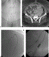
Example of progression of an enterocutaneous fistula progression to an enteroatmospheric fistula. A 66-year-old male presented with multiple small bowel resections due to adhesions from prior abdominal surgery requiring surgical bowel resection, subsequently complicated by enterocutaneous fistula. Postoperatively, the patient had clinically apparent enteral content from his laparotomy incision. (a) Initial computed tomography (CT) of the abdomen and pelvis with oral but no intravenous contrast demonstrates a high attenuation tract (arrow) adjacent to contrast-containing small bowel most consistent with enterocutaneous fistula. (b,c) Cannulation of this cutaneous tract and defect with water soluble contrast injection demonstrates opacification of adjacent bowel (arrows), suggestive of enterocutaneous fistula. (d) At 3-month follow- up, the patient’s laparotomy incision dehisced, resulting in a clinically apparent enteroatmospheric fistula (e) CT of the abdomen and pelvis with intravenous but not oral contrast demonstrates a defect of the bowel wall within a ventral hernia defect, with the sagittal reconstruction demonstrating the clinically apparent enteral content pooling in the wound’s defect (arrows).
Figure 1.
Figure 1.
Example of progression of an enterocutaneous fistula progression to an enteroatmospheric fistula. A 66-year-old male presented with multiple small bowel resections due to adhesions from prior abdominal surgery requiring surgical bowel resection, subsequently complicated by enterocutaneous fistula. Postoperatively, the patient had clinically apparent enteral content from his laparotomy incision. (a) Initial computed tomography (CT) of the abdomen and pelvis with oral but no intravenous contrast demonstrates a high attenuation tract (arrow) adjacent to contrast-containing small bowel most consistent with enterocutaneous fistula. (b,c) Cannulation of this cutaneous tract and defect with water soluble contrast injection demonstrates opacification of adjacent bowel (arrows), suggestive of enterocutaneous fistula. (d) At 3-month follow- up, the patient’s laparotomy incision dehisced, resulting in a clinically apparent enteroatmospheric fistula (e) CT of the abdomen and pelvis with intravenous but not oral contrast demonstrates a defect of the bowel wall within a ventral hernia defect, with the sagittal reconstruction demonstrating the clinically apparent enteral content pooling in the wound’s defect (arrows).
Figure 2.
Figure 2.
37-year-old male with Crohn’s disease status postsubtotal colectomy with J-pouch formation complicated by proctitis flares and persistent perianal fistulas. The patient presented with new midline fecal material draining from laparotomy incision. (a–c) Precontrast (a) and postcontrast (b) T1-weighted magnetic resonance (MR) axial images demonstrate a midline-enhancing inflammatory tract that restricts diffusion on diffusion-weighted imaging (c, arrow), consistent with the known midline enterocutaneous fistula. (d–g) More caudally to the midline enterocutaneous fistula above the urinary bladder an organized collection demonstrates enhancement (precontrast T1 [d] and postcontrast T1 [e]) and diffusion restriction (f) (arrows). The collection is also delineated on precontrast coronal T2-weighted sequence (g). This is all in keeping with a postoperative abscess–fistula complex in a patient with inflammatory bowel disease.
Figure 2.
Figure 2.
37-year-old male with Crohn’s disease status postsubtotal colectomy with J-pouch formation complicated by proctitis flares and persistent perianal fistulas. The patient presented with new midline fecal material draining from laparotomy incision. (a–c) Precontrast (a) and postcontrast (b) T1-weighted magnetic resonance (MR) axial images demonstrate a midline-enhancing inflammatory tract that restricts diffusion on diffusion-weighted imaging (c, arrow), consistent with the known midline enterocutaneous fistula. (d–g) More caudally to the midline enterocutaneous fistula above the urinary bladder an organized collection demonstrates enhancement (precontrast T1 [d] and postcontrast T1 [e]) and diffusion restriction (f) (arrows). The collection is also delineated on precontrast coronal T2-weighted sequence (g). This is all in keeping with a postoperative abscess–fistula complex in a patient with inflammatory bowel disease.
Figure 3.
Figure 3.
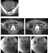
34-year-old morbidly obese woman presented with pelvic pain, fever, and leukocytosis. (a). Scout topogram of computed tomography (CT) of the abdomen and pelvis demonstrates the patient’s habitus. (b). Contrast-enhanced transaxial CT image demonstrates a large left adnexal fluid collection. This was initially managed with percutaneous drainage, which evacuated a large volume of purulent material (not pictured). (c,d) Follow-up catheter sinogram demonstrates opacification of the adjacent bowel loop (better appreciated on dynamic images) (e–g) Follow-up CT without intravenous contrast and with injection of indwelling drainage catheter demonstrates opacification of the adjacent loop of the sigmoid colon, consistent with fistulous communication.
Figure 3.
Figure 3.
34-year-old morbidly obese woman presented with pelvic pain, fever, and leukocytosis. (a). Scout topogram of computed tomography (CT) of the abdomen and pelvis demonstrates the patient’s habitus. (b). Contrast-enhanced transaxial CT image demonstrates a large left adnexal fluid collection. This was initially managed with percutaneous drainage, which evacuated a large volume of purulent material (not pictured). (c,d) Follow-up catheter sinogram demonstrates opacification of the adjacent bowel loop (better appreciated on dynamic images) (e–g) Follow-up CT without intravenous contrast and with injection of indwelling drainage catheter demonstrates opacification of the adjacent loop of the sigmoid colon, consistent with fistulous communication.
Figure 4.
Figure 4.
36-year-old female presented with Crohn’s disease and a history of multiple bowel resections and a chronic right lower quadrant abscess–fistula complex. (a) Initial contrast-enhanced transaxial computed tomography (CT) image demonstrates a large fluid collection; the patient was 2 weeks postoperative from an ileocecectomy. This was managed by percutaneous drainage. (b–f) On follow-up imaging, CT demonstrates that the fluid collection has resolved with an inflammatory tract about the course of the drainage catheter (b,c). Sinogram through existing catheter reveals a persistent fistulous tract to the adjacent bowel (d–f).
Figure 5.
Figure 5.
Steps for catheter management of abscess-fistula complexes. White arrow indicates direction of intestinal peristalsis, and black arrows depict enteric fluid leaking into the inflammatory walled-off abscess cavity. (a) Percutaneous abscess evacuation using two drainage catheters. (b) After the abscess cavity has decreased in size, a catheter is inserted in the loop of the bowel in the direction of peristalsis (white arrow) to cannulate the fistulous tract. One of the pigtail catheters draining the abscess has been removed as the cavity is resolving. As the residual abscess cavity is collapsed, the pigtail catheters are either downsized or removed. (c) By this time, there is a walled-off inflammatory tract from the intestine to the skin. The catheter cannulating the fistulous tract is removed after 48 hours of keeping it closed. (d) Abscess-fistula complex resolution. Reproduced with permission from (1): Ballard DH, Hamidian Jahromi A, Li AY, Vea R, Ahuja C, D’Agostino HB. Abscess-Fistula Complexes: A Systematic Approach for Percutaneous Catheter Management. J Vasc Interv Radiol. 2015;26:1363–1367; Elsevier, 2015.
Figure 6.
Figure 6.
53-year-old female with Crohn’s disease and a history of total abdominal colectomy (recently status: postloop ileostomy takedown and lysis of adhesions) presented with foul-smelling drainage from her surgical wound. (a,b) Transaxial contrast-enhanced computed tomography (CT) of the abdomen and pelvis shows a gas-filled, thickened tract extending from the inferior aspect of the patient’s surgical wound into the right lower quadrant toward a loop of the small bowel with surrounding fat stranding (arrows), consistent with an enterocutaneous fistula. (c–f) The enterocutaneous fistula was refractory to catheter drainage. Ten months after diagnosis, the patient underwent percutaneous closure of the fistula with a Biodesign plug. Part c shows a fistulous tract (yellow arrow) communicating with a loop of ileum (red arrow) in the right lower quadrant upon injection of contrast through the skin drainage site under fluoroscopy. Note the additional tract in the left lower quadrant (white arrow head) that is not in communication with bowel. Guidewire access and loading of the Biodesign plug into the fistulous tract are depicted in parts d to f. The arrows in parts e and f denote the plug device. The patient required surgical takedown of the enterocutaneous fistula and small bowel resection 2 months after this procedure.
Figure 6.
Figure 6.
53-year-old female with Crohn’s disease and a history of total abdominal colectomy (recently status: postloop ileostomy takedown and lysis of adhesions) presented with foul-smelling drainage from her surgical wound. (a,b) Transaxial contrast-enhanced computed tomography (CT) of the abdomen and pelvis shows a gas-filled, thickened tract extending from the inferior aspect of the patient’s surgical wound into the right lower quadrant toward a loop of the small bowel with surrounding fat stranding (arrows), consistent with an enterocutaneous fistula. (c–f) The enterocutaneous fistula was refractory to catheter drainage. Ten months after diagnosis, the patient underwent percutaneous closure of the fistula with a Biodesign plug. Part c shows a fistulous tract (yellow arrow) communicating with a loop of ileum (red arrow) in the right lower quadrant upon injection of contrast through the skin drainage site under fluoroscopy. Note the additional tract in the left lower quadrant (white arrow head) that is not in communication with bowel. Guidewire access and loading of the Biodesign plug into the fistulous tract are depicted in parts d to f. The arrows in parts e and f denote the plug device. The patient required surgical takedown of the enterocutaneous fistula and small bowel resection 2 months after this procedure.

References

    1. Ballard DH,Hamidian Jahromi A,Li AY,Vea R,Ahuja C, D’Agostino HB. Abscess-fistula complexes: a systematic approach for percutaneous catheter management. J Vasc Interv Radiol 2015;26(09): 1363–1367 - PubMed
    1. Lee JK, Stein SL. Radiographic and endoscopic diagnosis and treatment of enterocutaneous fistulas. Clin Colon Rectal Surg 2010;23(03):149–160 - PMC - PubMed
    1. Rahman FN, Stavas JM. Interventional radiologic management and treatment of enterocutaneous fistulae. J Vasc Interv Radiol 2015;26(01):7–19, quiz 20 - PubMed
    1. Kerlan RK Jr,Jeffrey RB Jr,Pogany AC,Ring EJ.Abdominalabscess with low-output fistula: successful percutaneous drainage. Radiology 1985;155(01):73–75 - PubMed
    1. Lee SS, Kim AY, Yang S-K, et al. Crohn disease of the small bowel: comparison of CT enterography, MR enterography, and small- bowel follow-through as diagnostic techniques. Radiology 2009; 251(03):751–761 - PubMed

LinkOut - more resources