

3D superimposition of craniofacial imaging-The utility of multicentre collaborations
- PMID: 31074129
- PMCID: PMC6660909
- DOI: 10.1111/ocr.12281
3D superimposition of craniofacial imaging-The utility of multicentre collaborations
Abstract
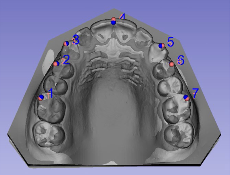
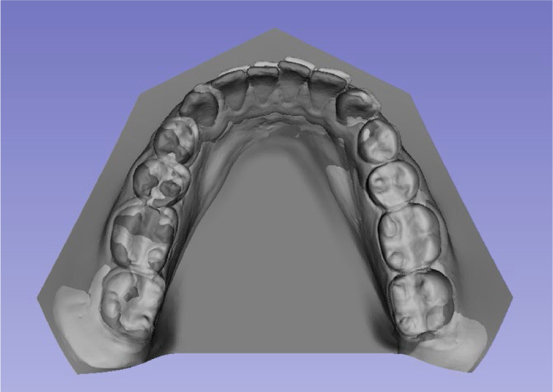
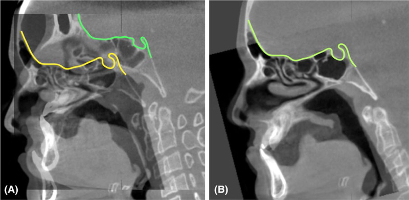
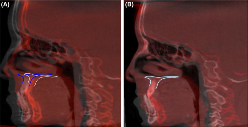
Clinical applications of 3D image registration and superimposition have contributed to better understanding growth changes and clinical outcomes. The use of 3D dental and craniofacial imaging in dentistry requires validate image analysis methods for improved diagnosis, treatment planning, navigation and assessment of treatment response. Volumetric 3D images, such as cone-beam computed tomography, can now be superimposed by voxels, surfaces or landmarks. Regardless of the image modality or the software tools, the concepts of regions or points of reference affect all quantitative of qualitative assessments. This study reviews current state of the art in 3D image analysis including 3D superimpositions relative to the cranial base and different regional superimpositions, the development of open source and commercial tools for 3D analysis, how this technology has increased clinical research collaborations from centres all around the globe, some insight on how to incorporate artificial intelligence for big data analysis and progress towards personalized orthodontics.
Keywords: 3D analysis; artificial intelligence; personalized orthodontics.
© 2019 John Wiley & Sons A/S. Published by John Wiley & Sons Ltd.
Figures
References
-
- Broadbent BH. A new x-ray technique and its application to orthodontia. Angle Orthod. 1931;1(2):45–66.
-
- Steiner CC. The use of cephalometrics as an aid to planning and assessing orthodontic treatment: report of a case. Am J Orthod. 1960;46(10):721–735.
-
- Mars M, James DR, Lamabadusuriya SP. The Sri Lankan Cleft Lip and Palate Project: the unoperated cleft lip and palate. Cleft Palate J. 1990;27(1):3–6. - PubMed
Publication types
MeSH terms
Grants and funding
LinkOut - more resources
Full Text Sources
