Review
. 2019 Oct 1;40(5):1424-1445.
doi: 10.1210/er.2018-00281.

Uterine Glands: Developmental Biology and Functional Roles in Pregnancy

Affiliations
Review

Uterine Glands: Developmental Biology and Functional Roles in Pregnancy

Andrew M Kelleher et al. Endocr Rev. .

Abstract

All mammalian uteri contain glands in the endometrium that develop only or primarily after birth. Gland development or adenogenesis in the postnatal uterus is intrinsically regulated by proliferation, cell-cell interactions, growth factors and their inhibitors, as well as transcription factors, including forkhead box A2 (FOXA2) and estrogen receptor α (ESR1). Extrinsic factors regulating adenogenesis originate from other organs, including the ovary, pituitary, and mammary gland. The infertility and recurrent pregnancy loss observed in uterine gland knockout sheep and mouse models support a primary role for secretions and products of the glands in pregnancy success. Recent studies in mice revealed that uterine glandular epithelia govern postimplantation pregnancy establishment through effects on stromal cell decidualization and placental development. In humans, uterine glands and, by inference, their secretions and products are hypothesized to be critical for blastocyst survival and implantation as well as embryo and placental development during the first trimester before the onset of fetal-maternal circulation. A variety of hormones and other factors from the ovary, placenta, and stromal cells impact secretory function of the uterine glands during pregnancy. This review summarizes new information related to the developmental biology of uterine glands and discusses novel perspectives on their functional roles in pregnancy establishment and success.


Figures

Figure 1.
Figure 1.
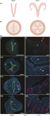
Uterine morphology, radial patterning, and postnatal development in mice and sheep. (a) Diagrams of ideal frontal sections of uterine types. The drawings cut the oviducts off near the uterotubal junctions, and the vagina just caudal to the cervix. Rodents (rats and mice) have a long duplex type of uterus with dual cervices. Sheep have a medium-length bicornuate type of uterus and a short uterine body and a single cervix. (b) Diagrams of ideal radial patterns of the uterine wall. The curved lines in the endometrium denote the tubular, coiled, and slightly branched glands that extend from the uterine lumen to the inner layer of myometrium. The mouse uterus lacks appreciable glands in the upper third of the endometrium on the mesometrial area of the uterus. The sheep uterus contains large number of glands in the intercaruncular areas of the endometrium, whereas the caruncles are glandless. (c) Immunofluorescent localization of forkhead box a2 (FOXA2) and keratin (KRT8) was performed for sections of the postnatal mouse uterus, whereas only FOXA2 was localized in the sheep uterus. Note the expression of FOXA2 in nascent and developing glands. Sections were counterstained with 4′,6-diamidino-2-phenylindole (DAPI) to visualize all nuclei. Scale bar, 100 μm (mouse) and 50 μm (sheep). Car, caruncle; M, myometrium; P, postnatal day; S, stroma.
Figure 2.
Figure 2.
Overview of uterine gland development in the mouse before puberty. (a) Dark-field images of the uterine epithelium isolated before (P5) and during genesis and budding of the glands from the LE (denoted by an asterisk). (b) Overview of uterine gland morphogenesis based on postnatal age, two-dimensional histology, and 3D imaging as proposed by Vue et al. (18).
Figure 3.
Figure 3.
Proposed source and actions of the WNT signaling system in postnatal uterine development in mice and sheep. (a) Schematic illustrating the canonical and noncanonical WNT signaling pathways and inhibition of those pathways by DKK and SFRP. Activation of the canonical signaling stimulates epithelial adhesion and proliferation as well as stromal cell proliferation in the intercaruncular endometrium. Activation of the noncanonical pathway would stimulate epithelial cell migration and movement. (b) Autocrine and paracrine actions of WNTs and their inhibitors in the neonatal mouse uterus. The upper panel displays immunofluorescent localization of forkhead box A2 (FOXA2) and keratin (KRT8) in the adult mouse uterus; note that FOXA2 is only expressed in the glands. In the lateral and antimesometrial (AM) stroma, WNTs expressed in the LE and stroma may have autocrine or paracrine actions on the LE, stroma, and/or GE to promote uterine adenogenesis. However, the WNT inhibitor DKK2 is expressed predominantly by the stroma of the mesometrial area (M) and myometrium that inhibits WNT signaling and thus inhibiting GE growth and development in the upper mesometrial area of the endometrium as well as the myometrium. Original magnification, ×10. (c) Autocrine and paracrine actions of WNTs and their inhibitors in the neonatal ovine uterus. The upper panel displays immunolocalization of FOXA2 in the uterus of a P14 ewe; note that FOXA2 is only expressed in the glands. In the intercaruncular endometrium, WNTs expressed in the LE (WNTs 5A, 7A, and 11) may have autocrine or paracrine actions on the LE or stroma, respectively. WNT2B is expressed only in the stroma and may have autocrine or paracrine actions on the stroma or the LE and GE, respectively. WNT5A is expressed predominantly by the GE and may have autocrine or paracrine actions on the GE or stroma, respectively. SFRP2 is expressed predominantly by the stroma of the caruncles as well as by the intercaruncular stroma between the tips of the glands and the inner circular layer of the myometrium. Binding of the FZD receptors for the WNTs on the epithelia and stroma inhibit epithelial growth and development into the caruncular areas of the endometrium as well as the myometrium. Scale bar, 50 μM. ABC, active β-catenin; Car, caruncle; CDH1, cadherin 1; DKK, dickkopf; DVL, dishevelled; FZD, frizzled receptor; GSK3B, glycogen synthase kinase 3β; LGR, leucine-rich repeat–containing G protein–coupled receptor; LPR, lipoprotein receptor-related protein; RSPO, roof plate–specific spondin; SFRP, secreted FZD-related protein; TCF, transcription factor.
Figure 4.
Figure 4.
Uterine glands secrete factors that impact pregnancy establishment in the mouse. In response to the nidatory surge in ovarian estrogen on day 4 of pregnancy, LIF is secreted from FOXA2-positive uterine glands, causing HBEGF expression for trophoblast attachment and adhesion to the LE. Following attachment, unknown gland-derived factors are involved in coordinating the removal of the LE (entosis) within the implantation chamber, allowing for direct contact between the trophoblast and decidualizing stromal cells (PTGS2). Uterine glands are involved in secondary stromal cell decidualization (SDZ), but the identification of factors involved in mediating those processes requires the establishment of new in vivo and in vitro model systems.
Figure 5.
Figure 5.
Hypothesis on the interrelationships of the ovarian corpus luteum, conceptus trophoblast, uterine glands, and decidual cells during early pregnancy in humans. See text for detailed description of hypotheses and supporting data. [Adapted from Burton GJ, Jauniaux E, Charnock-Jones DS. Human early placental development: potential roles of the endometrial glands. Placenta. 2007;28(Suppl A):S64–S69. This is an open access article distributed under the Creative Commons Attribution License, which permits unrestricted use, distribution, and reproduction in any medium, provided the original work is properly cited.]

References

    1. Gray CA, Bartol FF, Tarleton BJ, Wiley AA, Johnson GA, Bazer FW, Spencer TE. Developmental biology of uterine glands. Biol Reprod. 2001;65(5):1311–1323. - PubMed
    1. Cooke PS, Spencer TE, Bartol FF, Hayashi K. Uterine glands: development, function and experimental model systems. Mol Hum Reprod. 2013;19(9):547–558. - PMC - PubMed
    1. Spencer TE, Kelleher AM, Bartol FF. Development and function of uterine glands in domestic animals. Annu Rev Anim Biosci. 2019;7(1):125–147. - PubMed
    1. Bazer FW. Uterine protein secretions: relationship to development of the conceptus. J Anim Sci. 1975;41(5):1376–1382. - PubMed
    1. Roberts RM, Bazer FW. The functions of uterine secretions. J Reprod Fertil. 1988;82(2):875–892. - PubMed

Publication types