. 2019 Sep;34(9):1753-1766.
doi: 10.1002/jbmr.3756. Epub 2019 Jul 30.

Beclin1 Modulates Bone Homeostasis by Regulating Osteoclast and Chondrocyte Differentiation

Affiliations

Beclin1 Modulates Bone Homeostasis by Regulating Osteoclast and Chondrocyte Differentiation

Atsushi Arai et al. J Bone Miner Res. 2019 Sep.

Abstract

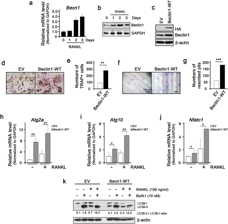
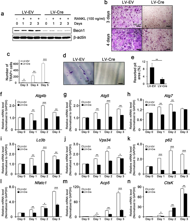
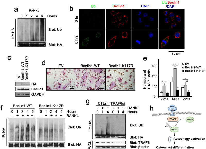
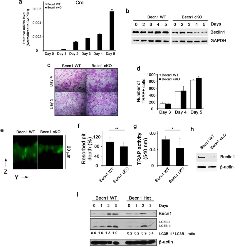
Autophagy (ATG), an important cellular recycling process whereby macromolecules or organelles are encapsulated by autophagosome and degraded upon merging with lysosome, has recently been shown to play an essential role in bone biology. However, the involvement of ATG in bone and bone-related cells remains unclear. Here, we show that Beclin1, an ATG-related protein involved in ATG initiation, plays a pivotal role in osteoclasts. ATG was activated during osteoclast differentiation in vitro. Beclin1 was enhanced and required for osteoclast differentiation. Mechanistically, we found that TRAF6-mediated ubiquitination of Beclin1 at K117, but not ULK1-mediated phosphorylation, is required for RANKL-stimulated osteoclast differentiation. In vivo, mice lacking Beclin1 in CstK-expressing cells exhibited an increased cortical bone thickness caused by impaired osteoclasts' function. Interestingly, these mice also exhibited diminished trabecular bone mass, which was associated with a defect in cartilage formation and chondrocyte differentiation. Collectively, our study highlights the functional importance of ATG in osteoclasts and chondrocytes, and identifies ATG as a potential therapeutic target for managing bone-related diseases. © 2019 American Society for Bone and Mineral Research.

Keywords: AUTOPHAGY; BECLIN1; CHONDROCYTES; OSTEOCLASTS; RANKL; UBIQUITINATION.


Figures

Figure 1
Figure 1. Beclin1 enhances RANKL-induced osteoclast differentiation.
(a) Real-time qPCR for expression of Becn1 mRNA during RANKL-induced osteoclast differentiation in RAW 264.7 cells. (b) Western blotting against Beclin1 and GAPDH during RANKL-induced osteoclast differentiation in RAW 264.7 cells. (c) Western blotting against HA, Beclin1, and β-actin in RAW 264.7 cells infected with retroviruses harboring empty vector (EV) or wildtype Beclin1 (Beclin1-WT). (d) TRAP staining following RANKL treatment on RAW 264.7 cells for 3 days. Bar = 100 μm. (e) Quantification of TRAP+ cells. (f) Dentin slice assay following RANKL treatment on RAW 264.7 cells for 10 days. Cells were stained with hematoxylin, removed the cells with cotton swabs, and examined under the light microscope. Bar = 100 μm. (g) Quantification of resorbed pits. (h,i,j) Real-time qPCR for expression of Atg2a, Atg10, and Nfatc1 in EV- or Beclin1-WT-expressing RAW 264.7 cells treated with RANKL. (k) Western blotting against LC3B and β-actin during RANKL-induced osteoclast differentiation in RAW 264.7 cells with or without BafA1.
Figure 2
Figure 2. Beclin1 is required for autophagy induction and osteoclast differentiation.
(a) Western blotting against Beclin1 and β-actin in RANKL/M-CSF-treated bone marrow macrophages (BMMs) isolated from Becn1 f/f mice and infected with lentiviruses harboring empty vector (LV-EV) or Cre (LV-Cre). (b) TRAP staining following RANKL/M-CSF treatment on EV- or Cre-expressing BMMs for 3 and 4 days. Bar = 100 μm. (c) Quantification of TRAP+ cells. (d) Dentin slice assay following RANKL/M-CSF treatment on EV- or Cre-expressing BMMs. Cells were stained with hematoxylin, removed with cotton swabs, and examined under the light microscope. Bar = 100 μm. (e) Quantification of resorbed pits. (f-k) Real-time qPCR for the expression of autophagy-related genes in EV- or Cre-expressing BMMs. (l,m,n) Real-time qPCR for the expression of osteoclast-related genes in EV- or Cre-expressing BMMs.
Figure 3
Figure 3. TRAF6-dependent ubiquitination of Beclin1 at K117 is required for RANKL-induced osteoclast differentiation.
(a) The ubiquitination assay in RANKL-treated RAW 264.7 cells in the indicated time points. (b) Fluorescent staining for ubiquitin (green) and Beclin1 (red) visualized using confocal microscopy. (c) Western blotting against HA, Beclin1, and β-actin in RAW 264.7 cells infected with retroviruses harboring empty vector (EV) or wildtype Beclin1 (Beclin1-WT), and ubiquitination incompetent mutant Beclin1 at K117 (Beclin1-K117R). (d) TRAP staining following RANKL treatment on RAW 264.7 cells for 3, 4 and 5 days. Bar = 200 μm. (e) Quantification of TRAP+ cells. (f) The ubiquitination assay in RANKL-treated RAW 264.7 cells expressing wildtype Beclin1 (Beclin1-WT) or ubiquitination incompetent mutant Beclin1 at K117 (Beclin1-K117R) in the indicated time points. (g) The ubiquitination assay in RANKL-treated RAW 264.7 cells expressing wildtype Beclin1 (Beclin1-WT) following transfection with CTL siRNA (CTLsi) and TRAF6 siRNA (TRAF6si). (h) A model for RANKL-induced autophagy activation via Beclin1 ubiquitination and osteoclast differentiation.
Figure 4
Figure 4. Becn1 cKO mice exhibited increased cortical bone thickness but diminished trabecular bone mass.
(a) Photographs of Becn1 WT and cKO mice. (b) Quantification of body weights at 6th month (n = 5 WT, n = 12 cKO, male). (c) Photographs of spleens from Becn1 WT and cKO mice. (d) Quantification of spleen at 6th month (n = 5 WT, n = 12 cKO, male) normalized to the body weights. (e) µCT of the femurs from Becn1 WT and cKO mice. (f) Quantification of femur lengths measured at 6th month (n = 5 WT, n = 12 cKO, male). (g) µCT of the femur cross-sections in Becn1 WT and cKO mice. (h) Quantification of femur cortical bone thickness measured at 6th month (n = 5 WT, n = 12 cKO, male). (i) µCT of the trabecular bone from Becn1 WT and cKO mice. Quantification of BV/TV (j), trabecular thickness (k), trabecular number (l), and trabecular spacing (m) measured at 6th month (n = 5 WT, n = 12 cKO, male). (n) H&E staining of the femur head. Bar = 200 µm. (o) TRAP staining in the femur trabecular bone. Bar = 100 µm. (p) Quantification of TRAP+ osteoclasts per bone perimeter. (q) TRAP staining in the femur cortical bone. Bar = 100 µm. (r) Quantification of the TRAP+ osteoclasts per bone perimeter.
Figure 5
Figure 5. Bone-resorbing function of osteoclasts is impaired in Becn1 cKO mice.
(a) qRT-PCR for Cre gene expression during osteoclast differentiation in BMMs isolated from Becn1 WT or cKO mice. (b) Western blotting for Beclin1 expression level during osteoclast differentiation. (c) TRAP staining following RANKL/M-CSF treatment on BMMs for 4 and 5 days. Bar = 100 μm. (d) Quantification of TRAP+ cells. (e) Dentin slice assay following RANKL/M-CSF treatment on BMMs isolated from Becn1 WT or cKO mice. Cells were removed with cotton swabs, and stained with FITC-wheat germ agglutinin (WGA). The resorbed pit depth on dentin slice was visualized in z stack by confocal microscopy. The image was reconstructed by Amira software. (f) Normalized resorbed pit depth excavated by Becn1 WT and cKO osteoclasts. Three different experiments (cell number = 100) were normalized to the average depth. (g) TRAP activity measurement from cell culture supernatant from dentin slices. (h) Western blotting for Beclin1 expression level from osteoclasts harvested from the dentin slices. (i) Western blotting against Beclin1, LC3B and β-actin during RANKL-induced osteoclast differentiation in BMMs from Becn1+/+ (WT) or Becn1+/− (Het) mice.
Figure 6
Figure 6. Chondrocyte differentiation is impaired in Becn1 cKO mice.
(a) Safranin O staining at the cartilage of Becn1 WT or cKO mice harvested at week 4, 8, and 24. (b) Quantification of the columnal zone at week 4 and 8. (c) Quantification of the hypertrophic zone at week 4 and 8. (d) Quantification of the total zone at week 4 and 8. (e) Immunohistochemical staining of Sox9, ColX, MMP13, and MMP9 at the cartilage. Bar = 100 µm. (f) Alcian blue staining of the chondrocytes isolated from the Becn1 WT and cKO mice at week 1, 2, and 3. (g) Quantification of Alcian blue staining.

Comment in

  • A role for autophagy in bone biology.
    Starling S. Starling S. Nat Rev Endocrinol. 2019 Aug;15(8):438-439. doi: 10.1038/s41574-019-0223-5. Nat Rev Endocrinol. 2019. PMID: 31142852 No abstract available.

References

    1. Katagiri T, Takahashi N. Regulatory mechanisms of osteoblast and osteoclast differentiation. Oral Dis May 2002;8(3):147–59. - PubMed
    1. Boyce BF, Rosenberg E, de Papp AE, Duong le T. The osteoclast, bone remodelling and treatment of metabolic bone disease. Eur J Clin Invest Dec 2012;42(12):1332–41. - PubMed
    1. Kobayashi K, Takahashi N, Jimi E, Udagawa N, Takami M, Kotake S, et al. Tumor necrosis factor alpha stimulates osteoclast differentiation by a mechanism independent of the ODF/RANKL-RANK interaction. J Exp Med Jan 17 2000;191(2):275–86. - PMC - PubMed
    1. Blair HC, Teitelbaum SL, Ghiselli R, Gluck S. Osteoclastic bone resorption by a polarized vacuolar proton pump. Science Aug 25 1989;245(4920):855–7. - PubMed
    1. Boya P, Reggiori F, Codogno P. Emerging regulation and functions of autophagy. Nat Cell Biol Jul 2013;15(7):713–20. - PMC - PubMed

Publication types

MeSH terms