. 2019 Jun 4;58(22):2617-2627.
doi: 10.1021/acs.biochem.9b00303. Epub 2019 May 23.

Structural, Kinetic, and Mechanistic Analysis of an Asymmetric 4-Oxalocrotonate Tautomerase Trimer

Structural, Kinetic, and Mechanistic Analysis of an Asymmetric 4-Oxalocrotonate Tautomerase Trimer

Bert-Jan Baas et al. Biochemistry. .

Abstract

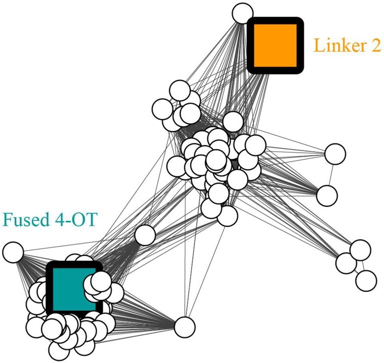
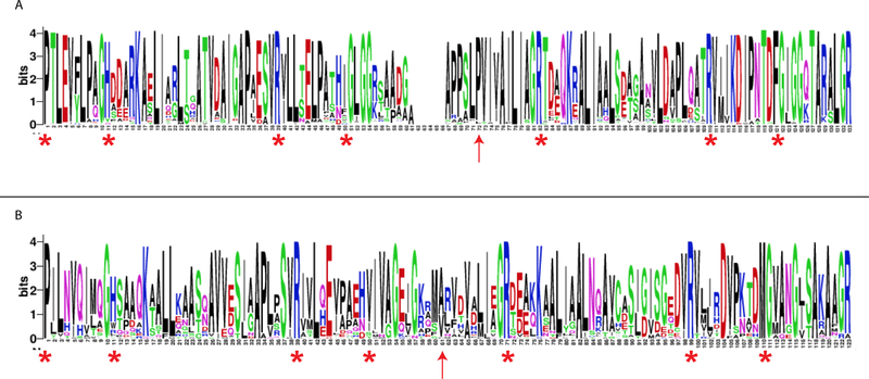
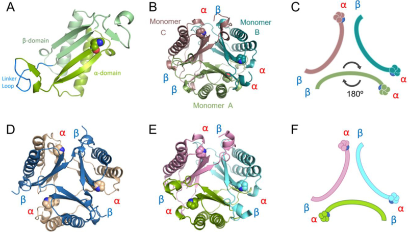
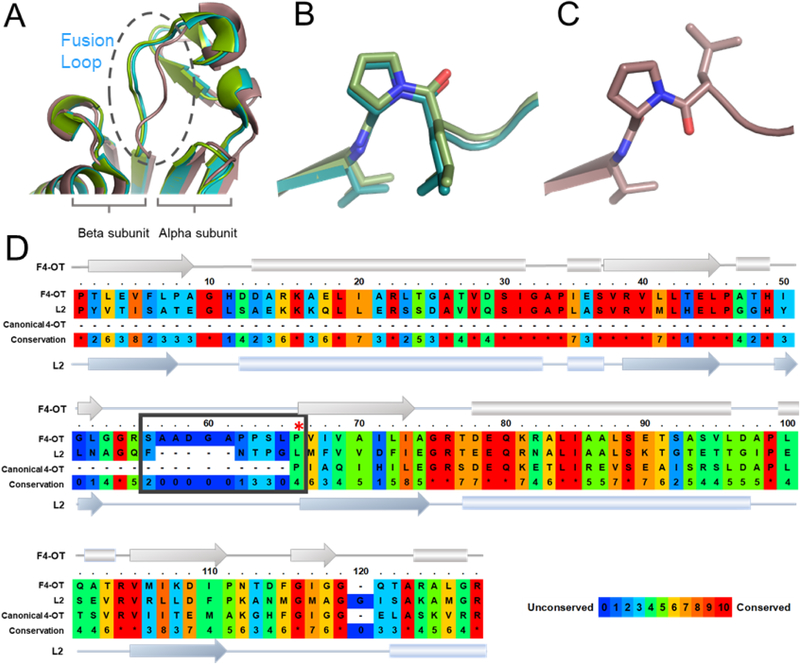
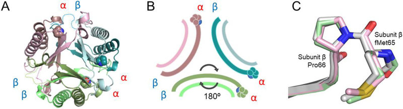
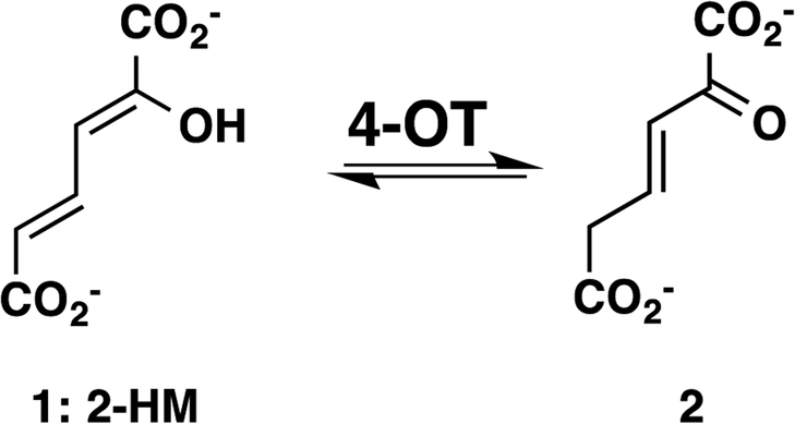
A 4-oxalocrotonate tautomerase (4-OT) trimer has been isolated from Burkholderia lata, and a kinetic, mechanistic, and structural analysis has been performed. The enzyme is the third described oligomer state for 4-OT along with a homo- and heterohexamer. The 4-OT trimer is part of a small subset of sequences (133 sequences) within the 4-OT subgroup of the tautomerase superfamily (TSF). The TSF has two distinct features: members are composed of a single β-α-β unit (homo- and heterohexamer) or two consecutively joined β-α-β units (trimer) and generally have a catalytic amino-terminal proline. The enzyme, designated as fused 4-OT, functions as a 4-OT where the active site groups (Pro-1, Arg-39, Arg-76, Phe-115, Arg-127) mirror those in the canonical 4-OT from Pseudomonas putida mt-2. Inactivation by 2-oxo-3-pentynoate suggests that Pro-1 of fused 4-OT has a low p Ka enabling the prolyl nitrogen to function as a general base. A remarkable feature of the fused 4-OT is the absence of P3 rotational symmetry in the structure (1.5 Å resolution). The asymmetric arrangement of the trimer is not due to the fusion of the two β-α-β building blocks because an engineered "unfused" variant that breaks the covalent bond between the two units (to generate a heterohexamer) assumes the same asymmetric oligomerization state. It remains unknown how the different active site configurations contribute to the observed overall activities and whether the asymmetry has a biological purpose or role in the evolution of TSF members.


Conflict of interest statement

The authors declare no competing financial interest.

Figures

Figure 1.
Figure 1.. Sequence similarity network showing the fused 4-OTs in the 4-OT subgroup.
The SNN is part of the Level 2 4-OT subgroup containing the canonical 4-OT (Figures S1 and S2). The SSN represents 133 sequences with characteristics shown in Figure 2. Nodes representing the characterized sequences for “fused 4-OT” and “Linker 2” are colored and labeled. Edges are drawn between two nodes only if the mean similarity between the two sequences is at least as significant as the bit score of 96 chosen to illustrate similarity relationships for this network.
Figure 2.
Figure 2.. Sequence logos for the fused 4-OT and Linker 2 clusters.
A) The sequence logo for the fused 4-OT cluster. The sequences of the “fused” 4-OTs (86 total) are characterized by two 4-OT-like sections joined in the middle by a linker-region, which results in a gap in the multiple sequence alignments (MSA). The red asterisks indicate the conserved features (described in the text) and the red arrow indicates a conserved proline residue (red arrow logo-position 72) that is 10 positions before the arginine at logo-position 82. This could represent the position of an N-terminal proline before fusion of two short 4-OTs in many of the sequences. B) The sequence logo for the Linker 2 cluster. The sequences show the conserved features (described in the text), but the positioning is not as striking. Moreover, there is not an obvious conservation of proline as the start of the C-terminal section (red arrow logo-position 61). The logos are graphical representations of multiple sequence alignments (MSAs) generated using WebLogo.
Figure 3.
Figure 3.
A) Ribbon diagram of one monomer of fused 4-OT. The α- and β-domains are labeled and colored in two shades of green. The linker loop is labeled and colored in bright blue. B) The fused 4-OT trimer, color coded and chains defined: monomer A in green (flipped monomer), monomer B in teal and monomer C in taupe. Interfaces defined between each monomer. C) Schematic of oligomerization arrangement of fused 4-OT. Color scheme is the same as in 3B and the position of Pro-1 is shown. D) Ribbon representation of the C. aurantiacus heterohexamer 4-OT with each interface defined. E) Ribbon representation of Linker 2 (a fused 4-OT) with each interface labeled. All interfaces are identical alpha-beta interfaces. F) In the same orientation as E, schematic of N-terminal β-strand containing Pro-1 of Linker 2, all monomers are symmetrical.
Figure 4.
Figure 4.
A) Superimposition of fused 4-OT monomers, close-up to the linker loop region. B) Stick view of L65/P66 motif overlay of monomers A and B showing cis-proline configuration. C) Stick view of L65/P66 motif monomer C showing Pro-66 in the trans configuration. D) Sequence alignment of fused 4-OT, Linker 2, and the canonical 4-OT (labeled as F4-OT, L2, and canonical 4-OT). The secondary structure representation is labeled in the same way. Linker loop is enclosed in a box. A red asterisk shows Pro-1 of canonical 4-OT aligned to Pro-66 of fused 4-OT. The PRALINE sequence alignment tool was used to make this figure.
Figure 5.
Figure 5.
A) Ribbon diagram of unfused 4-OT. Heterodimer A colored two shades of green, heterodimer B is colored two shades of teal and heterodimer C colored taupe/pink. The arrangement is an asymmetric trimer of dimers. The interfaces remain αα, αβ, and ββ, as labeled. The missing linker loop for each dimer is shown in dotted lines in corresponding colors. B) Schematic diagram of unfused 4-OT showing the flipped dimer and the corresponding interfaces. C) Overlay of three β-subunits of unfused 4-OT showing stick mode of the first three amino acids. The Leu-Pro sequence in the fused 4-OT is replaced by the initiating methionine followed by proline (Pro-66 in the fused 4-OT), in all β-subunits of the unfused 4-OT. The Met-Pro is in the trans configuration.
Scheme 1.
Scheme 1.
4-OT-catalyzed Reaction

References

    1. Whitman CP, Aird BA, Gillespie WR, and Stolowich NJ (1991) Chemical and enzymatic ketonization of 2-hydroxymuconate, a conjugated enol. J. Am. Chem. Soc 113, 3154–3162.
    1. Dagley S (1978) Pathways for the utilization of organic growth substrates, in The Bacteria: A Treatise on Structure and Function (Ornston LN and Sokatch JR, Eds), pp. 305–388, Academic Press, New York.
    1. Harayama S, Rekik M, Ngai K-L, and Ornston LN (1989) Physically associated enzymes produce and metabolize 2-hydroxy-2,4-dienoate, a chemically unstable intermediate formed in catechol metabolism via meta cleavage in Pseudomonas putida, J. Bacteriol 171, 6251–6258. - PMC - PubMed
    1. Stivers JT, Abeygunawardana C, Mildvan AS, Hajipour G, Whitman CP, and Chen LH (1996) Catalytic role of the amino-terminal proline in 4-oxalocrotonate tautomerase: affinity labeling and heteronuclear NMR studies. Biochemistry 35, 803–813. - PubMed
    1. Stivers JT, Abeygunawardana, Mildvan AS, Hajipour G, and Whitman CP (1996) 4-Oxalocrotonate tautomerase: pH dependences of catalysis and pKa values of active site residues. Biochemistry 35, 814–823. - PubMed

Publication types

Supplementary concepts

LinkOut - more resources