. 2019 Aug;33(8):9087-9099.
doi: 10.1096/fj.201802555R. Epub 2019 Jun 21.

ADP heptose, a novel pathogen-associated molecular pattern identified in Helicobacter pylori

Affiliations

ADP heptose, a novel pathogen-associated molecular pattern identified in Helicobacter pylori

Lennart Pfannkuch et al. FASEB J. 2019 Aug.

Erratum in

  • Corrigendum.
    [No authors listed] [No authors listed] FASEB J. 2020 Nov;34(11):15623. doi: 10.1096/fsb2.21084. Epub 2020 Oct 8. FASEB J. 2020. PMID: 33105917 Free PMC article. No abstract available.

Abstract

The gastric pathogen Helicobacter pylori activates the NF-κB pathway in human epithelial cells via the recently discovered α-kinase 1 TRAF-interacting protein with forkhead-associated domain (TIFA) axis. We and others showed that this pathway can be triggered by heptose 1,7-bisphosphate (HBP), an LPS intermediate produced in gram-negative bacteria that represents a new pathogen-associated molecular pattern (PAMP). Here, we report that our attempts to identify HBP in lysates of H. pylori revealed surprisingly low amounts, failing to explain NF-κB activation. Instead, we identified ADP-glycero-β-D-manno-heptose (ADP heptose), a derivative of HBP, as the predominant PAMP in lysates of H. pylori and other gram-negative bacteria. ADP heptose exhibits significantly higher activity than HBP, and cells specifically sensed the presence of the β-form, even when the compound was added extracellularly. The data lead us to conclude that ADP heptose not only constitutes the key PAMP responsible for H. pylori-induced NF-κB activation in epithelial cells, but it acts as a general gram-negative bacterial PAMP.-Pfannkuch, L., Hurwitz, R., Traulsen, J., Sigulla, J., Poeschke, M., Matzner, L., Kosma, P., Schmid, M., Meyer, T. F. ADP heptose, a novel pathogen-associated molecular pattern identified in Helicobacter pylori.

Keywords: ALPK1; LPS; NF-κB; PAMP; TIFA.


Conflict of interest statement

The authors thank A. Zamyatina (University of Natural Resources and Life Sciences, Vienna) for providing reference samples of ADP α-heptoses, Meike Sörensen (Max Planck Institute for Infection Biology, Berlin) for creation of plasmids, and Rike Zietlow (Max Planck Institute for Infection Biology, Berlin) for editing the manuscript. The authors declare no conflicts of interest.

Figures
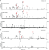
Figure 1
Figure 1
Detection of AEC-derivatized heptoses in H. pylori extracts by MALDI-TOF MSMS. A) HBP or extracts from H. pylori (graphite carbon eluate) were derivatized with AEC after acid hydrolysis, respectively. AEC heptose adducts were separated with UPLC and analyzed by MALDI-TOF MSMS. Shown are the MSMS spectra of the precursor ion m/z 485 [M+H]+ as the putative Schiff base–derived H7P-AEC adduct. Upper panel: Derivatization of H. pylori extracts, arrows and numbers in red: fragment ions (m/z 210, 223, 275 and 315) of the precursor ion m/z 485 [M+H]; middle: derivatization of HBP, lower: matrix control. B) Upper panel: MALDI-MSMS spectrum of the AEC-glycero-d-manno-heptose precursor ion (m/z 405 [M+H]+) derived from reductive amination of an H. pylori extract after acid hydrolysis with TFA; arrows and numbers in red: fragment ions (m/z 210, 223, 315) of the precursor ion m/z 405 [M+H]. Lower: matrix control.
Figure 2
Figure 2
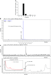
Identification of a novel NF-κB–stimulating compound in H. pylori lysates. A) AGS NF-κB luciferase reporter cells were incubated with H. pylori fractions separated by ion-pairing reversed-phase UPLC. Fractions were mixed with transfection reagent (lipofectamine 2000) and then added to NF-κB luciferase reporter cells for 3 h. Induction of NF-κB was determined by measurement of luciferase activity. Treatment with lipofectamine 2000 was used as negative control. Induction is shown as fold change compared with a negative control. Representative result of 2 repeated runs is shown. B) H. pylori extract was spiked with β-HBP. Shown are the chromatograms in SIR mode for m/z 369 (black line) and m/z 618 (negative mode) (blue, dashed line). Corresponding fractions in A are given below. C) H. pylori extract (carbon graphite eluate) was separated by UPLC. Shown are the chromatograms in SIR mode for m/z 618 (upper panel) and the UV absorbance at 259 nm (lower panel).
Figure 3
Figure 3
NF-κB–stimulating compound in H. pylori lysates is β-ADP heptose. A) MALDI-TOF MSMS spectra of ADP heptoses, m/z 618.2 [M-H]; corresponding fragment ions are labeled 1) Reference spectrum: α-d-ADP heptose; 2) β-d-ADP heptose from H1P or AMP morpholidate synthesis; 3) β-d-ADP heptose from H1P/HldE enzymatic synthesis; 4) β-d-ADP heptose from purified H. pylori extracts; 5) Matrix control. B) AGS NF-κB luciferase reporter cells were stimulated with chemically synthesized ADP-α-d-heptose and β-d-ADP heptose, and d- and l-glycero isomers as well as with enzymatically generated β-d-ADP heptose in d and l forms in the presence of a transfection reagent for 3 h at concentrations of 10 µM. Induction is shown as fold change compared with untreated control. Data are means ± sem of at least 2 independent replicates.
Figure 4
Figure 4
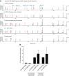
GmhB is essential for β-ADP heptose synthesis in H. pylori and necessary for NF-κB activation. A) Solid phase extracts from lysates of H. pylori WT and isogenic mutants ΔgmhA, ΔgmhB, and ΔrfaE were run over a Waters HSS T3 reversed-phase UPLC. Shown are the chromatograms in SIR mode for m/z 618 (negative mode). B) AGS cells were infected with H. pylori WT and indicated mutants at multiplicity of infection 100 for 3 h and analyzed for IL-8 induction by qRT-PCR. Data show relative induction compared with respective uninfected control. NI, not infected; Rel. relative. Data represent means ± sem of 2 independent replicates.
Figure 5
Figure 5
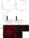
β-ADP heptose activates the ALPK1-TIFA axis at nanomolar concentrations. A, B) Treatment of AGS NF-κB luciferase reporter cells with β-d-ADP heptose and HBP in presence (A) or absence (B) of a transfection reagent for 3 h at indicated concentrations. Induction is shown as fold-change compared with an untreated control. Data represent means ± sem of 3 independent experiments. C) ALPK1−/− and TIFA−/− cells were stimulated with β-d-ADP heptose at a concentration of 10 µM in presence or absence of transfection reagent for 3 h or left untreated and analyzed for IL-8 induction by qRT-PCR. Data represent means ± sem of at least 2 independent replicates. D) AGS cells stably overexpressing tdTomato-TIFA were left untreated (control), treated with β-d-ADP heptose or HBP in presence or absence of a transfection reagent or infected with P12 WT at MOI 100 for 3 h. Formation of TIFAsomes was analyzed by confocal microscopy. Ns, not significant; NT, untreated. Scale bar, 20 µm. ***P ≤ 0.001, ****P ≤ 0.0001, Student’s t test.

References

    1. Janeway C. A., Jr (1989) Approaching the asymptote? Evolution and revolution in immunology. Cold Spring Harb. Symp. Quant. Biol. 54, 1–13 - PubMed
    1. Medzhitov R. (2007) Recognition of microorganisms and activation of the immune response. Nature 449, 819–826 - PubMed
    1. Baccala R., Gonzalez-Quintial R., Lawson B. R., Stern M. E., Kono D. H., Beutler B., Theofilopoulos A. N. (2009) Sensors of the innate immune system: their mode of action. Nat. Rev. Rheumatol. 5, 448–456 - PubMed
    1. DiDonato J. A., Mercurio F., Karin M. (2012) NF-κB and the link between inflammation and cancer. Immunol. Rev. 246, 379–400 - PubMed
    1. Newton K., Dixit V. M. (2012) Signaling in innate immunity and inflammation. Cold Spring Harb. Perspect. Biol. 4, a006049 - PMC - PubMed

MeSH terms

LinkOut - more resources