

Microglia: Lifelong patrolling immune cells of the brain
- PMID: 31075285
- PMCID: PMC6599472
- DOI: 10.1016/j.pneurobio.2019.04.003
Microglia: Lifelong patrolling immune cells of the brain
Abstract
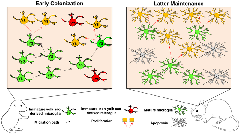
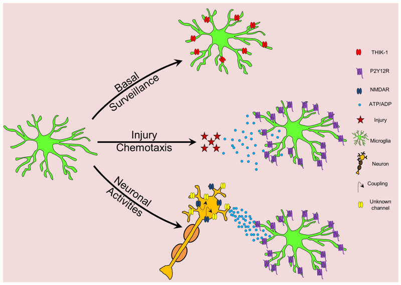
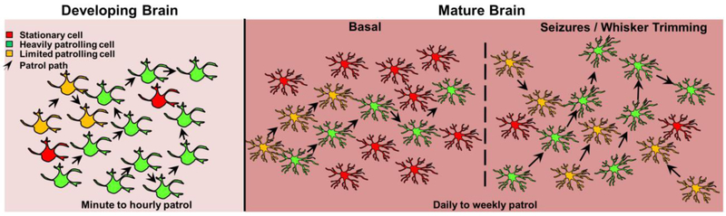
Microglial cells are the predominant parenchymal immune cell of the brain. Recent evidence suggests that like peripheral immune cells, microglia patrol the brain in health and disease. Reviewing these data, we first examine the evidence that microglia invade the brain mesenchyme early in embryonic development, establish residence therein, proliferate and subsequently maintain their numbers throughout life. We, then, summarize established and novel evidence for microglial process surveillance in the healthy and injured brain. Finally, we discuss emerging evidence for microglial cell body dynamics that challenge existing assumptions of their sessile nature. We conclude that microglia are long-lived immune cells that patrol the brain through both cell body and process movements. This recognition has significant implications for neuroimmune interactions throughout the animal lifespan.
Keywords: Epilepsy; Microglia; Microglial landscape; Neuroimmune interaction; Surveillance.
Copyright © 2019 Elsevier Ltd. All rights reserved.
Conflict of interest statement
Figures
References
-
- Ajami B, Bennett JL, Krieger C, McNagny KM, Rossi FM, 2011. Infiltrating monocytes trigger EAE progression, but do not contribute to the resident microglia pool. Nature neuroscience 14, 1142–1149. - PubMed
-
- Ajami B, Bennett JL, Krieger C, Tetzlaff W, Rossi FM, 2007. Local self-renewal can sustain CNS microglia maintenance and function throughout adult life. Nat Neurosci 10, 1538–1543. - PubMed
-
- Alliot F, Godin I, Pessac B, 1999. Microglia derive from progenitors, originating from the yolk sac, and which proliferate in the brain. Brain Res Dev Brain Res 117, 145–152. - PubMed
-
- Askew K, Li K, Olmos-Alonso A, Garcia-Moreno F, Liang Y, Richardson P, Tipton T, Chapman MA, Riecken K, Beccari S, Sierra A, Molnar Z, Cragg MS, Garaschuk O, Perry VH, Gomez-Nicola D, 2017. Coupled Proliferation and Apoptosis Maintain the Rapid Turnover of Microglia in the Adult Brain. Cell Rep 18, 391–405. - PMC - PubMed
Publication types
MeSH terms
Grants and funding
LinkOut - more resources
Full Text Sources
Medical
