. 2019 Jun 18;50(6):1401-1411.e4.
doi: 10.1016/j.immuni.2019.04.003. Epub 2019 May 7.

Inflammasome Activation Triggers Blood Clotting and Host Death through Pyroptosis

Affiliations

Inflammasome Activation Triggers Blood Clotting and Host Death through Pyroptosis

Congqing Wu et al. Immunity. .

Abstract

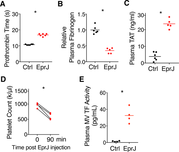
Inflammasome activation and subsequent pyroptosis are critical defense mechanisms against microbes. However, overactivation of inflammasome leads to death of the host. Although recent studies have uncovered the mechanism of pyroptosis following inflammasome activation, how pyroptotic cell death drives pathogenesis, eventually leading to death of the host, is unknown. Here, we identified inflammasome activation as a trigger for blood clotting through pyroptosis. We have shown that canonical inflammasome activation by the conserved type III secretion system (T3SS) rod proteins from Gram-negative bacteria or noncanonical inflammasome activation by lipopolysaccharide (LPS) induced systemic blood clotting and massive thrombosis in tissues. Following inflammasome activation, pyroptotic macrophages released tissue factor (TF), an essential initiator of coagulation cascades. Genetic or pharmacological inhibition of TF abolishes inflammasome-mediated blood clotting and protects against death. Our data reveal that blood clotting is the major cause of host death following inflammasome activation and demonstrate that inflammasome bridges inflammation with thrombosis.

Keywords: DIC; GSDMD; LPS; caspase; coagulation; inflammasome; macrophage; pyroptosis; sepsis; tissue factor.


Conflict of interest statement

DECLARATION OF INTERESTS

D.K. is an employee of Genentech Inc. The other authors declare no competing interests.

Figures

Figure 1.
Figure 1.. Administration of EprJ in vivo Induces Systemic Coagulation (A-E)
Mice (C57BL/6J) were injected intravenously with PBS (Ctrl) or EprJ (300 ng LFn-EprJ plus 3 μg PA per mouse). Blood were collected 90 minutes after PBS or EprJ injection. Prothrombin time (A), plasma fibrinogen concentrations (B), plasma TAT concentrations (C), total platelet count before and after EprJ injection (D), and TF activity in plasma microvesicles (MVs) (E) were measured. Solid circles represent individual mice, crossbars represent group mean. n = 4–6 for all experimental groups. One asterisk, P < 0.01 (Student’s t-test, unpaired for A-C, E, paired for D). See also Figure S1
Figure 2.
Figure 2.. Caspase-1 is Required for EprJ-induced Blood Coagulation and Lethality (A)
C57BL/6J mice (WT) or Casp1/11 deficient mice were injected intravenously with washed platelets isolated from the C57BL/6-Tg(CAG-EGFP)1Osb/J mice (GFP) and an Alexa 568-labled (Red) anti-fibrin monoclonal antibody (59D8), followed by intravenous injection of EprJ. Thrombus formation in cremaster vasculatures was monitored using an intravital microscopy. Images were acquired at the same location, 15 min and 60 min after EprJ injection. Scale bar denotes 50 μm. Data are representative of 3 independent experiments (biological replicates). (B) C57BL/6J mice (WT) or Casp1/11 deficient mice were injected intravenously with EprJ. After 90 minutes, mice were euthanized and perfused with PBS then perfusion-fixed with 10% formalin under physiological pressure for 45 minutes. Liver sections were immunostained with the anti-fibrin monoclonal antibody (59D8). Wild type mice, but not Casp1/11−/− mice, showed fibrin deposition in liver (arrows). Scale bar denotes 50 μm. Data are representative of 3 independent experiments (biological replicates). (C) C57BL/6J mice (WT) or Casp1/11 deficient mice were injected intravenously with EprJ. After 90 minutes, mice were euthanized and tissues were isolated. Fibrin in the tissue lysates was detected by Immunoblot with the anti-fibrin monoclonal antibody (59D8). Data are representative of 3 independent experiments (biological replicates). (D-F) C57BL/6J mice (WT), Casp1/11 deficient mice, Casp11 deficient mice, and TLR4 deficient mice were injected intravenously with PBS or EprJ. Blood were collected 90 minutes after PBS or EprJ injection. Prothrombin time (D), plasma fibrinogen concentrations (E), and plasma TAT concentrations (F) were measured. Error bars denote SEM. n = 4–6 for all experimental groups. Two asterisks, P < 0.01 (two-way ANOVA with Holm-Sidak multiple comparisons). (G) C57BL/6J mice (WT) or Casp1/11 deficient mice were injected intravenously with a lethal dose of EprJ. Kaplan-Meier survival plots for mice challenged with EprJ. n = 12–15. Three asterisks, P < 0.01 versus WT [Log-rank (Mantel-Cox) test]. See also Figure S1
Figure 3.
Figure 3.. GSDMD-dependent Pyroptosis is Essential to Inflammasome-driven Coagulation and Lethality (A)
C57BL/6J (WT) or GSDMD deficient mice were injected intravenously with EprJ. After 90 minutes, mice were euthanized and tissues were isolated. Fibrin in the tissue lysates was detected by Immunoblot with the anti-fibrin monoclonal antibody (59D8). Data are representative of 3 independent experiments (biological replicates). (B-D) C57BL/6J mice (WT) or GSDMD deficient mice were injected intravenously with PBS (Ctrl) or EprJ. Blood were collected 90 minutes after PBS or EprJ injection. Prothrombin time (B), plasma fibrinogen concentrations (C), and plasma TAT concentrations (D) were measured. Error bars denote SEM. n = 4–6 for all experimental groups. Two asterisks, P < 0.01 (two-way ANOVA with Holm-Sidak multiple comparisons). (E and F) C57BL/6J mice (WT) or GSDMD deficient mice were injected intravenously with PBS (Ctrl) or BsaK or PrgJ. Blood were collected 90 minutes after the injection. Prothrombin time (E), plasma TAT concentrations (F) were measured. Error bars denote SEM. n = 5 for all experimental groups. Two asterisks, P < 0.01 (two-way ANOVA with Holm-Sidak multiple comparisons). (G and H) C57BL/6J mice (WT) or GSDMD deficient mice were injected intravenously with a lethal dose of EprJ or BsaK. Kaplan-Meier survival plots for mice challenged with EprJ. n = 8–12. Three asterisks, P < 0.01 versus WT [Log-rank (Mantel- Cox) test]. See also Figure S2–S4
Figure 4.
Figure 4.. Macrophages Pyroptosis Plays a Critical Role in Inflammasome-induced Coagulation by Promoting the Releasing of TF-positive Microvesicles (A-C)
C57BL/6J mice were injected intravenously with control liposomes (Lipo) or clodronate- containing liposomes (Cldn) 24 hours prior to intravenous injection of PBS (Ctrl) or EprJ. Blood were collected 90 minutes after PBS or EprJ injection. Prothrombin time (A), plasma fibrinogen concentrations (B), and plasma TAT concentrations (C) were measured. Error bars denote SEM. n = 4–6 for all experimental groups. Two asterisks, P < 0.01 (two-way ANOVA with Holm- Sidak multiple comparisons). (D) C57BL/6J mice were injected intravenously with control liposomes (Lipo) or clodronate-containing liposomes (Cldn) 24 hours prior to intravenous injection of a lethal dose EprJ. Three asterisks, P < 0.01 [Log-rank (Mantel-Cox) test]. (E) BMDMs from C57BL/6J (WT), Casp1/11 deficient, Casp11 deficient mice, and TLR4 deficient mice were incubated with various T3SS rod proteins (100 ng/ml LFn-rod protein plus 1 μg/ml PA) for 45 minutes. TF and p20 caspase-1 in the supernatant were detected by fluorescent Immunoblot. Cell culture supernatants were precipitated to concentrate protein prior to Immunoblot. (F) BMDMs from C57BL/6J (WT) and TLR4 deficient mice were incubated with various T3SS rod proteins for 45 minutes. TF and p20 caspase-1 in the supernatant were detected by fluorescent Immunoblot. (G) BMDMs from C57BL/6J (WT) and GSDMD deficient mice were incubated with EprJ for 45 minutes. TF and p20 caspase-1 in the supernatant were detected by fluorescent Immunoblot. Glycine (+) designates addition of 5 mM glycine as osmoprotectant to prevent pyroptosis-associated membrane rupture. (H) THP-1 cells were incubated with T3SS needle protein EprI (1 ug/ml LFn-EprI plus 1μg/ml PA) for 45 minutes. Cell culture supernatants were precipitated to concentrate protein prior to Immunoblot analysis. Data are representative of 3 independent experiments for all the Immunoblot (biological replicates). (I) THP-1 were incubated with EprI for 45 minutes. Microvesicles (MVs) were isolated from supernatant and TF activity was measured. n = 4–6 for all experimental groups (biological replicates). One asterisk, P < 0.01 (Student’s t-test, unpaired). (J) Plasma MV TF activity. C57BL/6J (WT), Casp1/11 deficient, and GSDMD deficient mice were injected intravenously with EprJ. Blood were collected 90 minutes after EprJ injection. Microvesicles (MVs) were isolated from blood and TF activity was measured. Error bars denote SEM. n = 4–6 for all experimental groups. Four asterisks, P < 0.01 (one-way ANOVA with Holm-Sidak multiple comparisons). See also Figure S5
Figure 5.
Figure 5.. Tissue Factor Inhibition Protects Against Inflammasome-induced Coagulation and Lethality (A-C)
Pharmacological inhibition of TF. C57BL/6J mice were injected intravenously with a rat IgG or a rat anti-moue TF neutralizing antibody 1H1 (8 mg/kg). After 2 hours, the mice were injected intravenously with EprJ. Blood were collected 90 minutes after EprJ injection. Prothrombin time (A) and plasma TAT concentrations (B) were measured. Error bars denote SEM. n = 4–5 for all experimental groups. One asterisk, P < 0.01 (Student’s t-test, unpaired). (C) C57BL/6J mice were injected intravenously with a rat IgG or 1H1 (8 mg/kg). After 2 hours, the mice were injected intravenously with a lethal dose of EprJ. Three asterisks, P < 0.01 [Log-rank (Mantel-Cox) test]. (D-F) Inducible TF deficient mice was generated by crossing B6.Cg-Tg(UBC-cre/ERT2)1Ejb/J Cre transgenic mice with TF floxed mice. TF deficient mice or wild type littermates were injected intravenously with EprJ. Blood were collected 90 minutes after EprJ injection. Prothrombin time (D) and plasma TAT concentrations (E) were measured. Error bars denote SEM. n = 4–5 for all experimental groups. One asterisk, P < 0.01 (Student’s t-test, unpaired). (F) TF deficient mice or wild type littermates were injected with a lethal dose of EprJ. n = 8. Three asterisks, P < 0.01 [Log-rank (Mantel-Cox) test]. See also Figure S5
Figure 6.
Figure 6.. Noncanonical inflammasome activation by LPS triggers blood coagulation (A-D)
C57BL/6J mice (WT), Casp11 deficient, and TLR4 deficient mice were injected intraperitoneally with 4 mg/kg poly(I:C) for priming. After 8 hours, the mice were injected intraperitoneally with PBS (Ctrl) or 50 mg/kg LPS. Blood were collected 4 hours after injection of PBS or LPS injection. Prothrombin time (A), plasma fibrinogen concentrations (B), plasma TAT concentrations (C), and total platelet count before and after LPS injection (D) were measured. Error bars denote SEM. n = 4–6 for all experimental groups. Two asterisks, P < 0.01 (two-way ANOVA with Holm-Sidak multiple comparisons). (E) BMDMs from C57BL/6J (WT), Casp11 deficient mice, TLR4 deficient, and GSDMD deficient mice were incubated with poly(I:C) (1 μg/ml). After 5 hours, the cells were transfected with PBS (Ctrl) or LPS (2 μg/ml). Cell culture supernatants were precipitated to concentrate protein prior to Immunoblot. Data are representative of 3 independent experiments (biological replicates). (F-I) C57BL/6J mice (WT) or GSDMD deficient mice were injected intraperitoneally with 4 mg/kg poly(I:C) for priming. After 8 hours, the mice were injected intraperitoneally with PBS (Ctrl) or 50 mg/kg LPS. Blood were collected 4 hours after injection of PBS or LPS injection. Prothrombin time (F), plasma fibrinogen concentrations (G), plasma TAT concentrations (H), and total platelet count before and after LPS injection (I) were measured. Error bars denote SEM. n = 4–6 for all experimental groups. Two asterisks, P < 0.01 (two-way ANOVA with Holm-Sidak multiple comparisons). (J) C57BL/6J mice were injected intravenously with control liposomes (Lipo) or clodronate- containing liposomes (Cldn) 24 hours prior to intraperitoneal injection of 4 mg/kg poly(I:C). After 6 hours of poly(I:C) injection, the mice injected with a lethal dose of LPS. Three asterisks, P < 0.01 [Log-rank (Mantel-Cox) test]. (K) Model of coagulation triggered by canonical and noncanonical inflammasome activation. See also Figure S6

Comment in

References

    1. Aglietti RA, Estevez A, Gupta A, Ramirez MG, Liu PS, Kayagaki N, Ciferri C, Dixit VM, and Dueber EC (2016). GsdmD p30 elicited by caspase-11 during pyroptosis forms pores in membranes. Proc Natl Acad Sci U S A 113, 7858–7863. - PMC - PubMed
    1. Angus DC, and van der Poll T (2013). Severe sepsis and septic shock. N Engl J Med 369, 840–851. - PubMed
    1. Aziz M, Jacob A, Yang WL, Matsuda A, and Wang P (2013). Current trends in inflammatory and immunomodulatory mediators in sepsis. J Leukoc Biol 93, 329–342. - PMC - PubMed
    1. Bach R, Nemerson Y, and Konigsber W (1981). Purification and characterization of bovine tissue factor. The Journal of Biological Chemistry 256, 8324–8331. - PubMed
    1. Boucher D, Monteleone M, Coll RC, Chen KW, Ross CM, Teo JL, Gomez GA, Holley CL, Bierschenk D, Stacey KJ, et al. (2018). Caspase-1 self-cleavage is an intrinsic mechanism to terminate inflammasome activity. J Exp Med 215, 827–840. - PMC - PubMed

Publication types

MeSH terms