Meta-Analysis
. 2019 Aug;138(2):309-326.
doi: 10.1007/s00401-019-02020-0. Epub 2019 May 10.

Second-generation molecular subgrouping of medulloblastoma: an international meta-analysis of Group 3 and Group 4 subtypes

Affiliations
Meta-Analysis

Second-generation molecular subgrouping of medulloblastoma: an international meta-analysis of Group 3 and Group 4 subtypes

Tanvi Sharma et al. Acta Neuropathol. 2019 Aug.

Abstract

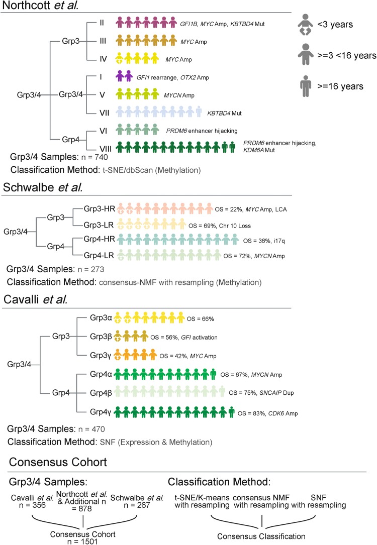
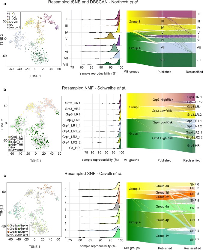
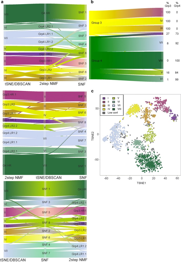
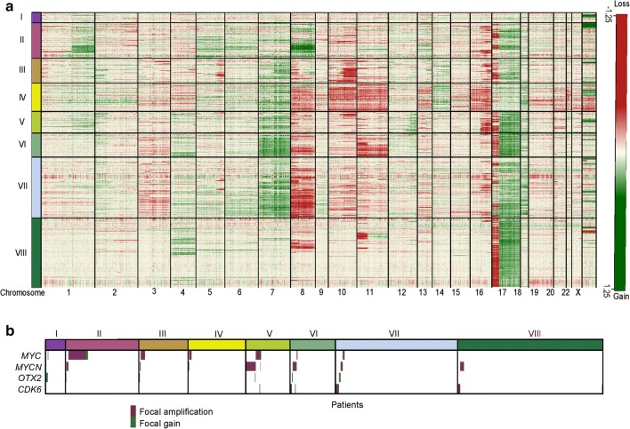
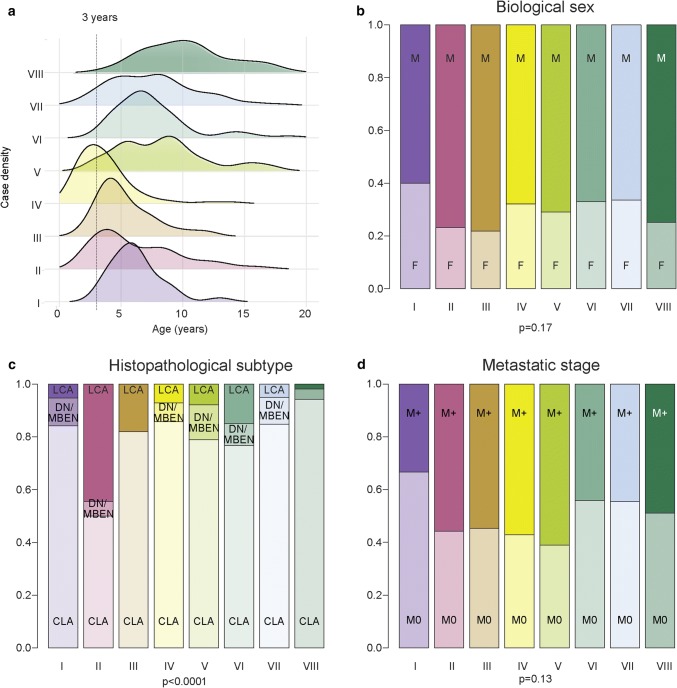
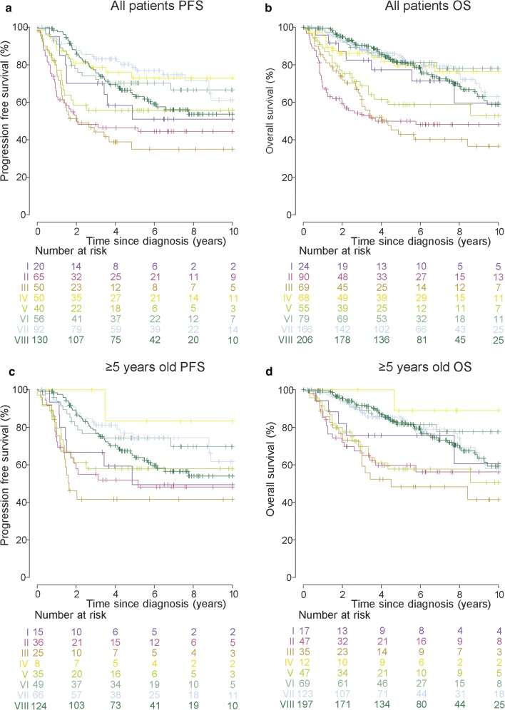
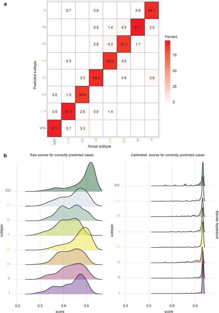
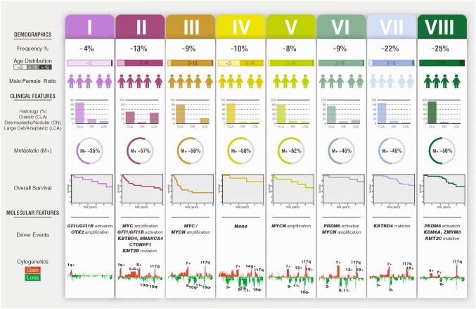
In 2012, an international consensus paper reported that medulloblastoma comprises four molecular subgroups (WNT, SHH, Group 3, and Group 4), each associated with distinct genomic features and clinical behavior. Independently, multiple recent reports have defined further intra-subgroup heterogeneity in the form of biologically and clinically relevant subtypes. However, owing to differences in patient cohorts and analytical methods, estimates of subtype number and definition have been inconsistent, especially within Group 3 and Group 4. Herein, we aimed to reconcile the definition of Group 3/Group 4 MB subtypes through the analysis of a series of 1501 medulloblastomas with DNA-methylation profiling data, including 852 with matched transcriptome data. Using multiple complementary bioinformatic approaches, we compared the concordance of subtype calls between published cohorts and analytical methods, including assessments of class-definition confidence and reproducibility. While the lowest complexity solutions continued to support the original consensus subgroups of Group 3 and Group 4, our analysis most strongly supported a definition comprising eight robust Group 3/Group 4 subtypes (types I-VIII). Subtype II was consistently identified across all component studies, while all others were supported by multiple class-definition methods. Regardless of analytical technique, increasing cohort size did not further increase the number of identified Group 3/Group 4 subtypes. Summarizing the molecular and clinico-pathological features of these eight subtypes indicated enrichment of specific driver gene alterations and cytogenetic events amongst subtypes, and identified highly disparate survival outcomes, further supporting their biological and clinical relevance. Collectively, this study provides continued support for consensus Groups 3 and 4 while enabling robust derivation of, and categorical accounting for, the extensive intertumoral heterogeneity within Groups 3 and 4, revealed by recent high-resolution subclassification approaches. Furthermore, these findings provide a basis for application of emerging methods (e.g., proteomics/single-cell approaches) which may additionally inform medulloblastoma subclassification. Outputs from this study will help shape definition of the next generation of medulloblastoma clinical protocols and facilitate the application of enhanced molecularly guided risk stratification to improve outcomes and quality of life for patients and their families.

Keywords: Biomarkers; Medulloblastoma; Meta-analysis; Methylation; Subtypes.


Figures

Fig. 1
Fig. 1
Summary of ‘second-generation’ medulloblastoma subgrouping of Grp3/4 medulloblastoma. a For each component study, the reported subtypes of Grp3/4 are shown, alongside incidence, age where possible, methodology, and major study findings. For each individual study, the most frequent subtype was scaled to 14 human figures; less frequently occurring subtypes were scaled from there. LR low risk, HR high risk. The analytical approach employed in a unified Grp3/4 cohort in this study is outlined in b. t-SNE t-stochastic neighbor embedding, NMF non-negative matrix factorization, SNF similarity network fusion
Fig. 2
Fig. 2
Consensus clustering identifies substructure within Grp3/4 medulloblastoma. Each row shows the results of applying t-SNE (a), NMF (b), and SNF (c), respectively, to the Grp3/4 cohort (n = 1501). For each row, a common t-SNE visualization is shown in the first panel, which depicts study-specific samples with their original study-specific subtype designation. The samples not included in a study were annotated as empty ‘NA’ in the t-SNEs (marked as empty circles). The samples that could not be assigned to a concordant subtype for a corresponding technique were annotated as ‘Low conf’ (marked in grey). In the second column, the reproducibility of the identified sample subtype is shown. Density plots show the distribution of sample reproducibility from consensus clustering by subtype for the selected number of clusters for each approach—t-SNE, NMF, and SNF. In the third column, Sankey plots demonstrate the relationship of the published subtypes to the assigned subtypes when clustered as part of a larger cohort. c The SNF results are from analysis of DNA-methylation data only
Fig. 3
Fig. 3
Inter-technique comparisons identify eight subtypes within Grp3/4 medulloblastoma. a Sankey plots show the relationship of subtype calls from each technique to those from other techniques. The same information is repeated three times for the Sankey plot; each plot shows a different technique in the center, to enable inter-technique relationships to be identified. b Sankey plot shows relationship between subtype calls and Grp 3 and 4 subgroup membership. c t-SNE visualizing subtype calls of 1501 Grp3/4 samples at weighted standard deviation > 0.25 with 15,335 most variable probes. Samples assigned to the same subtype from ≥ 2 techniques were assigned to that subtype. Unassignable samples were annotated as ‘Low conf’. DBSCAN density-based spatial clustering algorithm, used to identify subtypes after t-SNE dimension reduction. Two-step NMF refers to the two stages of NMF analysis; five robust subtypes were identified in the first stage. In the second stage, NMF consensus clustering was applied to each of the five subtypes in isolation
Fig. 4
Fig. 4
Subtypes have distinct copy-number profiles and significant enrichments of oncogene amplification. a CNV heatmap generated from raw conumee calls on methylation data for each subtype across all chromosomes revealed subtype-specific cytogenetic aberrations. Gains are shown in green, losses in red. b oncoplot shown summarizes the type and incidence of aberrations for MYC, MYCN, OTX2, and CDK6. Focal amplifications are shown in burgundy, focal gains are shown green
Fig. 5
Fig. 5
Clinico-pathological associations of Grp3/4 subtypes. a Subtypes show distinct age distributions. Density plot shows age distribution for each subtype. bd Barplots show incidence of major clinico-pathological features (sex, histopathology, and metastatic stage, respectively) across subtypes. b No difference in distribution of biological sex is evident. c Subtype II is significantly enriched for large-cell anaplastic histology. d No difference in metastatic disease (i.e., M0 vs M1+ disease) between subgroups. p values shown are derived from Chi squared tests of enrichment performed across all subtypes
Fig. 6
Fig. 6
Grp3/4 subtypes have distinct survival outcomes. Kaplan–Meier plots are shown for PFS/OS for all samples with available survival data (a, b), and to avoid confounding by patients treated with infant protocols, a filtered cohort comprised of patients aged ≥ 5 years at diagnosis (c, d). The at-risk table below the Kaplan–Meier plots (I–VIII) shows the number of patients at risk at specific times after diagnosis (0–10 years in 2 year intervals)
Fig. 7
Fig. 7
Molecular classifier for Grp3/4 subtype identification. a Confusion matrix shows relationship between predicted and actual subtype calls for the Grp3/4 cohort. The minimum reproducibility across subtypes was > 90%. b Density plots show raw and calibrated scores from classifier (refer to supplementary methods for detailed information). After calibration, the prediction accuracy increased, with an overall error rate of 4.6%
Fig. 8
Fig. 8
Summary of molecular subtypes of Grp3/4 medulloblastoma. The major demographic, clinico-pathological, and molecular features of the concordant subtypes are summarized. Mutation data were derived solely from Northcott et al. [18]. For histology, CLAS classic, DN desmoplastic nodular, LCA large-cell anaplastic. M+ (%) = metastatic (i.e., M1+) frequency. Overall survival shows subtype-specific survival in years. Cytogenetic gains are shown in red, losses in green

References

    1. Archer TC, Ehrenberger T, Mundt F, Gold MP, Krug K, Mah CK, Mahoney EL, Daniel CJ, LeNail A, Ramamoorthy D, et al. Proteomics, post-translational modifications, and integrative analyses reveal molecular heterogeneity within medulloblastoma subgroups. Cancer Cell. 2018;34(3):396–410. doi: 10.1016/j.ccell.2018.08.004. - DOI - PMC - PubMed
    1. Capper D, Jones DTW, Sill M, Hovestadt V, Schrimpf D, Sturm D, Koelsche C, Sahm F, Chavez L, Reuss DE, et al. DNA methylation-based classification of central nervous system tumours. Nature. 2018;555:469–474. doi: 10.1038/nature26000. - DOI - PMC - PubMed
    1. Cavalli FMG, Remke M, Rampasek L, Peacock J, Shih DJH, Luu B, Garzia L, Torchia J, Nor C, Morrissy AS, et al. Intertumoral heterogeneity within medulloblastoma subgroups. Cancer Cell. 2017;31(6):737–754. doi: 10.1016/j.ccell.2017.05.005. - DOI - PMC - PubMed
    1. Cho YJ, Tsherniak A, Tamayo P, Santagata S, Ligon A, Greulich H, Berhoukim R, Amani V, Goumnerova L, Eberhart CG, et al. Integrative genomic analysis of medulloblastoma identifies a molecular subgroup that drives poor clinical outcome. J Clin Oncol. 2011;29:1424–1430. doi: 10.1200/JCO.2010.28.5148. - DOI - PMC - PubMed
    1. Clifford SC, Lusher ME, Lindsey JC, Langdon JA, Gilbertson RJ, Straughton D, Ellison DW. Wnt/Wingless pathway activation and chromosome 6 loss characterize a distinct molecular sub-group of medulloblastomas associated with a favorable prognosis. Cell Cycle. 2006;5:2666–2670. doi: 10.4161/cc.5.22.3446. - DOI - PubMed

Publication types

MeSH terms