

A case of giant cell tumor of the breast, clinically suspected as malignant breast tumor
- PMID: 31076887
- PMCID: PMC6510746
- DOI: 10.1186/s40792-019-0635-4
A case of giant cell tumor of the breast, clinically suspected as malignant breast tumor
Abstract
Background: Giant cell tumor (GCT) of the breast is scarce. We report a case of GCT of the breast which was suspected as a malignant breast tumor.
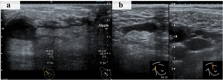
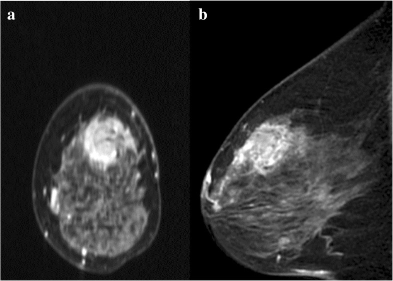
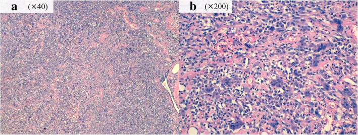
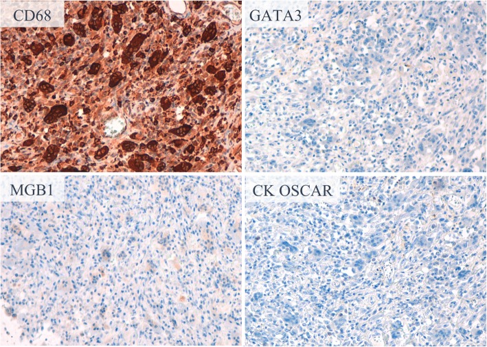
Case presentation: A 74-year-old woman noticed a tender lump in her right breast. We suspected a malignant tumor spreading widely with axillary lymph node metastasis on clinical examination and imaging. Histological evaluation of the biopsy tissue revealed a tumor composed the proliferation of oval to spindle-shaped cells and multinucleated giant cells without malignant epithelial cells. The tumor cells stained positively for CD68 and negatively for estrogen receptor, progesterone receptor, and human epidermal growth factor receptor 2. The pathological findings suggested GCT, and fine needle aspiration biopsy for the axillary lymph node was negative. However, there was a gap between the clinical presentation, such as a tender mass suggesting rapid growth and multiple lymphadenopathies, and the pathological presentation of biopsy, which made us hesitate to conclude GCT as the final preoperative diagnosis. We could not rule out the possibility of malignant tumors with OGCs before surgery. We performed mastectomy and sentinel lymph node biopsy according to a surgical procedure for node-negative breast cancer with a wide ductal spread. The resected tissue histologically showed the same findings to the biopsy tissue. The definitive diagnosis of GCT of the breast was given, because the tumor lacked epithelial components, marked cellular atypia, and pleomorphism.
Conclusions: GCT of the breast occasionally pretends as breast malignant tumors. Complete tumor resection should be performed for local control and the definitive diagnosis.
Keywords: Breast tumor; Giant cell tumor; Giant cell tumor of soft tissue; Metaplastic carcinoma.
Conflict of interest statement
Ethics approval and consent to participate
Ethical approval: All procedures performed involving human participants were in accordance with the ethical standards of the Institutional Review Board of Aichi Cancer Center and with the 1964 Helsinki declaration and its later amendments.
Informed consent: Informed consent was obtained from the patient for this publication.
Consent for publication
Not applicable.
Competing interests
The authors declare that they have no competing interests.
Publisher’s Note
Springer Nature remains neutral with regard to jurisdictional claims in published maps and institutional affiliations.
Figures
References
-
- Oliveira A. Giant cell tumour of soft tissues. In: Fletcher CDM, Bridge JA, Hogendoorn PCW, eds MF, editors. World Health Organization classification of tumours of soft tissue and bone. Lyon: IARC Press; 2013.
LinkOut - more resources
Full Text Sources
Research Materials
Miscellaneous
