. 2019 May 10;19(1):100.
doi: 10.1186/s12862-019-1429-9.

Functional diversification of horizontally acquired glycoside hydrolase family 45 (GH45) proteins in Phytophaga beetles

Affiliations

Functional diversification of horizontally acquired glycoside hydrolase family 45 (GH45) proteins in Phytophaga beetles

André Busch et al. BMC Evol Biol. .

Abstract

Background: Cellulose, a major polysaccharide of the plant cell wall, consists of β-1,4-linked glucose moieties forming a molecular network recalcitrant to enzymatic breakdown. Although cellulose is potentially a rich source of energy, the ability to degrade it is rare in animals and was believed to be present only in cellulolytic microbes. Recently, it has become clear that some animals encode endogenous cellulases belonging to several glycoside hydrolase families (GHs), including GH45. GH45s are distributed patchily among the Metazoa and, in insects, are encoded only by the genomes of Phytophaga beetles. This study aims to understand both the enzymatic functions and the evolutionary history of GH45s in these beetles.

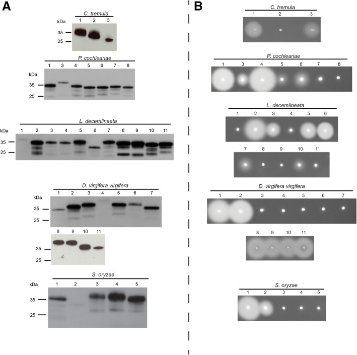
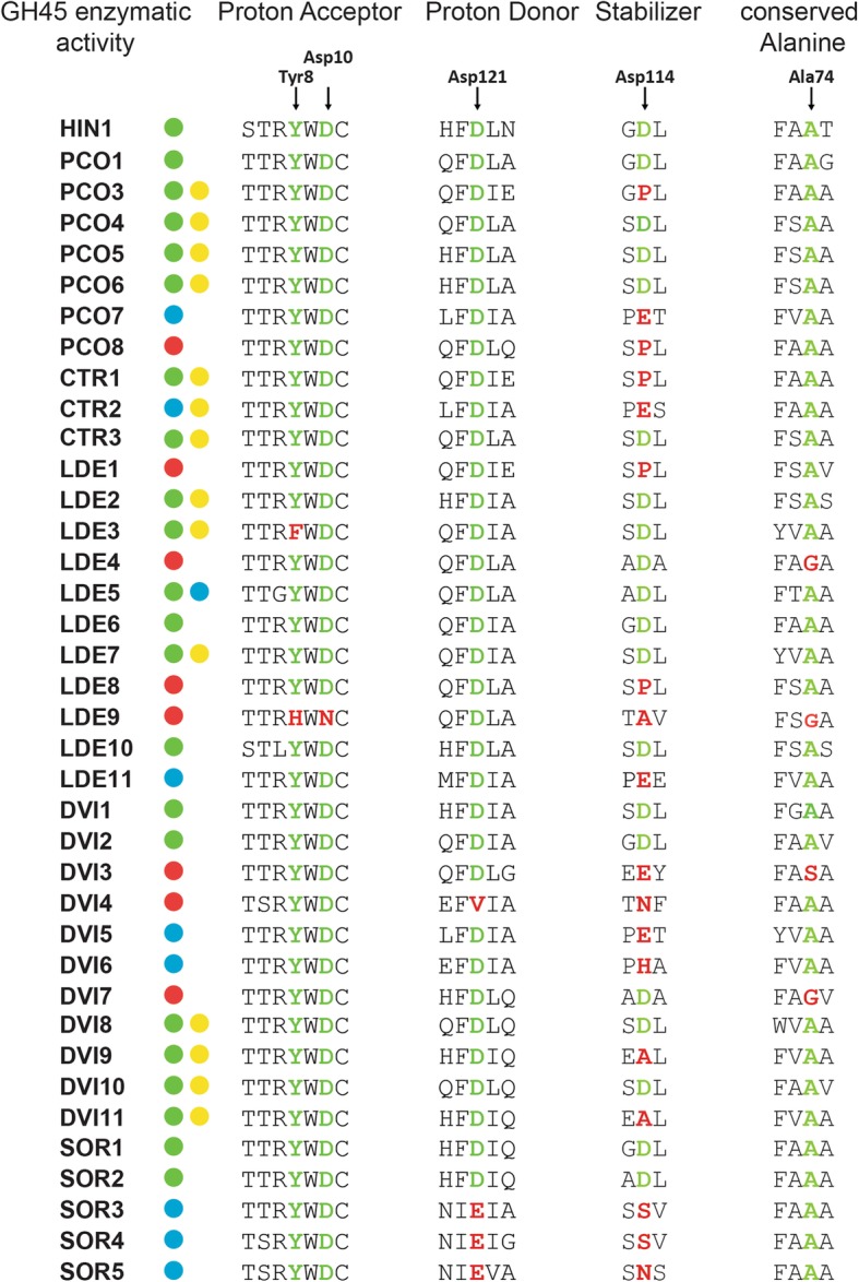
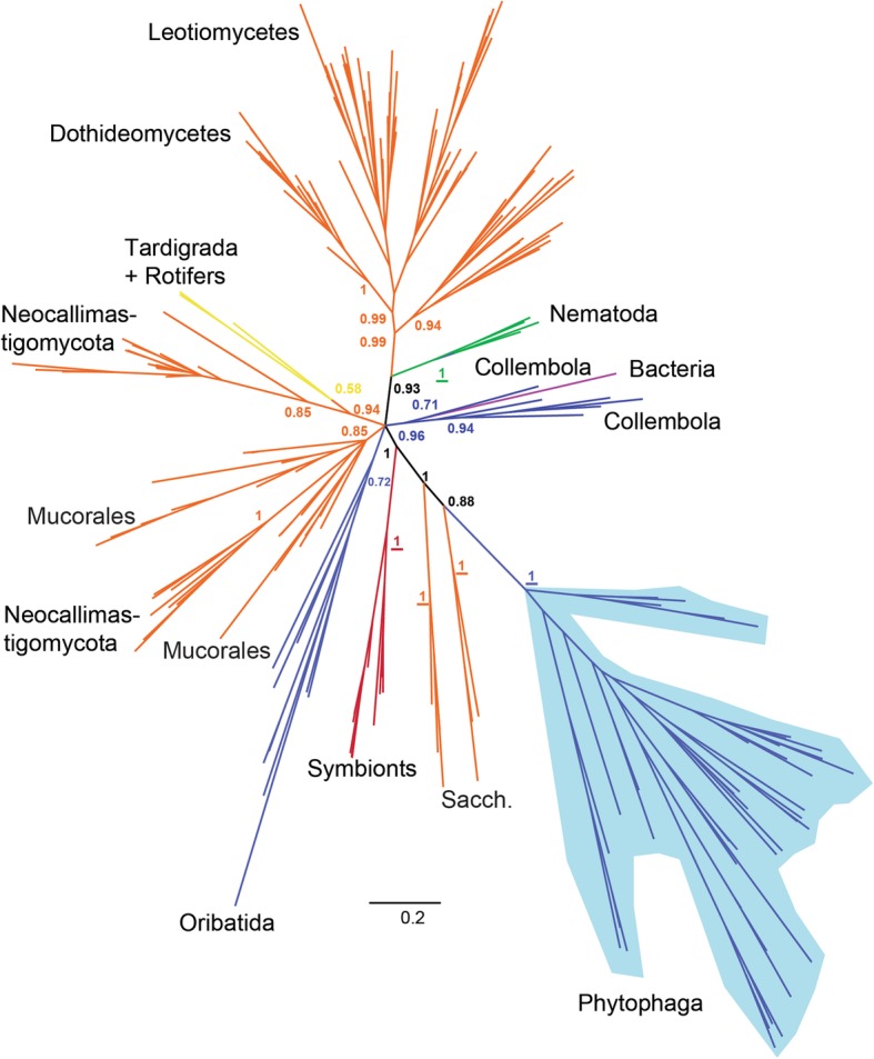
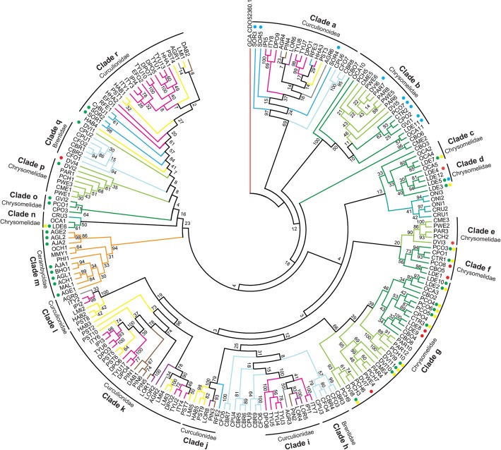
Results: To this end, we biochemically assessed the enzymatic activities of 37 GH45s derived from five species of Phytophaga beetles and discovered that beetle-derived GH45s degrade three different substrates: amorphous cellulose, xyloglucan and glucomannan. Our phylogenetic and gene structure analyses indicate that at least one gene encoding a putative cellulolytic GH45 was present in the last common ancestor of the Phytophaga, and that GH45 xyloglucanases evolved several times independently in these beetles. The most closely related clade to Phytophaga GH45s was composed of fungal sequences, suggesting this GH family was acquired by horizontal gene transfer from fungi. Besides the insects, other arthropod GH45s do not share a common origin and appear to have emerged at least three times independently.

Conclusion: The rise of functional innovation from gene duplication events has been a fundamental process in the evolution of GH45s in Phytophaga beetles. Both, enzymatic activity and ancestral origin suggest that GH45s were likely an essential prerequisite for the adaptation allowing Phytophaga beetles to feed on plants.

Keywords: Cellulase; Chrysomeloidea; Curculionoidea; GH45; Horizontal gene transfer; Xyloglucanase.


Conflict of interest statement

Ethics approval and consent to participate

Not applicable

Consent for publication

Not applicable

Competing interests

The authors declare that they have no competing interests.

Publisher’s Note

Springer Nature remains neutral with regard to jurisdictional claims in published maps and institutional affiliations.

Figures

Fig. 1
Fig. 1
Western blot and CMC-based agarose-diffusion assay of target GH45 proteins. a) Western blot of target recombinant enzymes expressed in frame with a V5/(His)6 after heterologous expression in insect Sf9 cells. After 72 h, crude culture medium of transfected cells was harvested and analyzed by Western blotting using an anti-V5 HRP-coupled antibody. b) Crude culture medium of transfected cells was applied to an agarose-diffusion assay containing 0.1% CMC. Activity halos were revealed after 16 h incubation at 40 °C using Congo red. Numbers above Western blot and agarose-diffusion assays correspond to the respective species of GH45s depicted in Additional file 1: Table S1
Fig. 2
Fig. 2
GH45 amino acid alignment of the catalytic residues. We used a GH45 sequence of Humicola insulens (HIN1) as a reference sequence (Accession: 2ENG_A) [37]. According to HIN1, we chose to investigate the catalytic residues (ASP10 and ASP121) as well as a conserved tyrosine (TYR8) of the catalytic binding site, a crucial substrate-stabilizing amino acid (ASP114) and an essential conserved alanine (ALA74). Arrows indicate amino acid residues under investigation. If the respective amino acid residue is highlighted in green, it is retained in comparison to the reference sequence; otherwise it is highlighted in red. GH45 enzymatic activity was color-coded based on the respective substrate specificity (green dots = endo-β-1,4-glucanase, blue dots = endo-β-1,4-xyloglucanase, yellow dots = (gluco)mannanase, red dots = no detected activity)
Fig. 3
Fig. 3
Global phylogeny encompassing GH45 proteins from various taxa. Bayesian-based phylogenetic analysis of GH45 sequences. 264 GH45 sequences of microbial and metazoan origin were initially collected (see Methods), and their redundancy was eliminated at 90% sequence similarity, resulting in a total of 201 sequences. Posterior probability values are given at crucial branches. If values are depicted in bold, the same branch appeared in the corresponding maximum likelihood analysis (see Additional file 1: Figures S6 and S7). If underlined, the maximum likelihood node was highly supported (bootstrap values > 75). Detailed sequence descriptions including accession numbers are given in Additional file 1: Table S2. Arthropoda are represented in blue, fungi in orange, protists in red, bacteria in purple, Nematoda in green and other Metazoa in yellow. Sacch. = Saccharomycetales fungi; Neocallim. = Neocallimastigaceae fungi
Fig. 4
Fig. 4
Phylogenetic relationships of Phytophaga-derived GH45s. A maximum-likelihood-inferred phylogeny of the predicted amino acid sequences of beetle-derived GH45s was performed. Bootstrap values are indicated at corresponding branches. Information on sequences and their accession number are given in Additional file 1: Table S2. Dots indicate GH45s characterized to date and are color-coded based on their activity: green = endo-β-1,4-glucanases; blue = endo-β-1,4-xyloglucanases; yellow = (gluco)mannanases; red = no activity detected. Color coding in reference to the respective subfamily of Curculionoidea: pink = Scolytinae (Curculionidae); brown = Entiminae (Curculionidae); purple = Cyclominae (Curculionidae); gray = Curculioninae (Curculionidae); yellow = Molytinae (Curculionidae); light blue = Brentinae (Brentidae); dark blue = Dryophthorinae (Curculionidae). Color coding in reference to the respective subfamily of Chrysomeloidea: dark green = Chrysomelinae (Chrysomelidae); light green = Galerucinae (Chrysomelidae); orange = Lamiinae (Cerambycidae); cyan = Cassidinae (Chrysomelidae)

References

    1. Chang MM, Chou T. Y. C., Tsao, G. T.: Structure, pretreatment and hydrolysis of cellulose In: Bioenergy Advances in Biochemical Engineering, vol 20. Springer, Berlin, Heidelberg; 1981. 15–42.
    1. Ruel K, Nishiyama Y, Joseleau JP. Crystalline and amorphous cellulose in the secondary walls of Arabidopsis. Plant Sci. 2012;193-194:48–61. - PubMed
    1. Cosgrove DJ. Re-constructing our models of cellulose and primary cell wall assembly. Curr Opin Plant Biol. 2014;22:122–131. - PMC - PubMed
    1. Knox JP. Revealing the structural and functional diversity of plant cell walls. Curr Opin Plant Biol. 2008;11(3):308–313. - PubMed
    1. Saxena IM, Brown RM. A perspective on the assembly of cellulose-synthesizing complexes: possible role of korrigan and microtubules in cellulose synthesis in plants. In: Brown RM, Saxena IM, editors. Cellulose: molecular and structural biology. Dordrecht: Springer; 2007. pp. 169–181.

Publication types

LinkOut - more resources