. 2019 May 10;20(1):14.
doi: 10.1186/s12865-019-0293-0.

Identification and characterisation of transient receptor potential melastatin 2 and CD38 channels on natural killer cells using the novel application of flow cytometry

Affiliations

Identification and characterisation of transient receptor potential melastatin 2 and CD38 channels on natural killer cells using the novel application of flow cytometry

Cassandra Balinas et al. BMC Immunol. .

Abstract

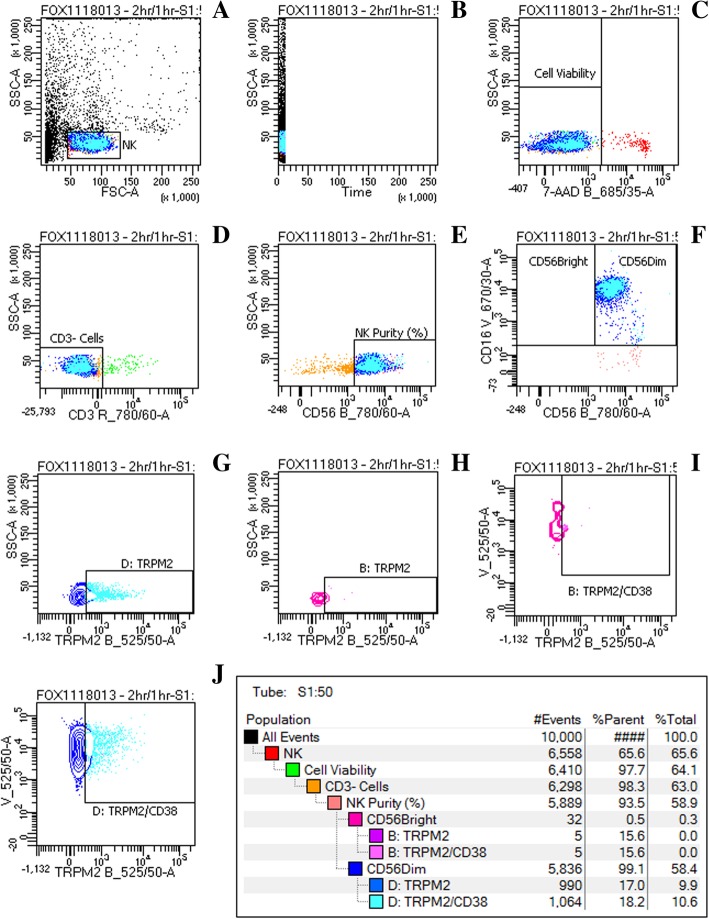
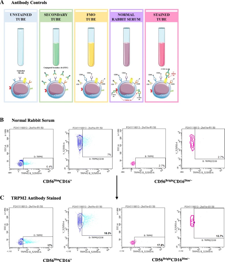
Background: Natural Killer (NK) cells are effector lymphocytes of the innate immune system and are subclassed into CD56BrightCD16Dim/- and CD56DimCD16+ NK cells. Intracellular calcium (Ca2+) is fundamental to regulate a number of intracellular signalling pathways and functions in NK cells, which are essential in mediating their natural cytotoxic function. Transient receptor potential melastatin 2 (TRPM2) is a Ca2+-permeable non-selective cation channel that possesses a critical role in calcium-dependent cell signalling to maintain cellular homeostasis. TRPM2 and CD38 protein surface expression has yet to be determined on NK cells using flow cytometry. Characterisation of TRPM2 has been previously identified by in vivo models, primarily using methods such as genetic remodification, immunohistochemistry and whole cell electrophysiology. The aim of this study was to develop an in vitro methodology to characterise TRPM2 and CD38 surface expression on NK cell subsets using an antibody that has not been previously applied using flow cytometry.

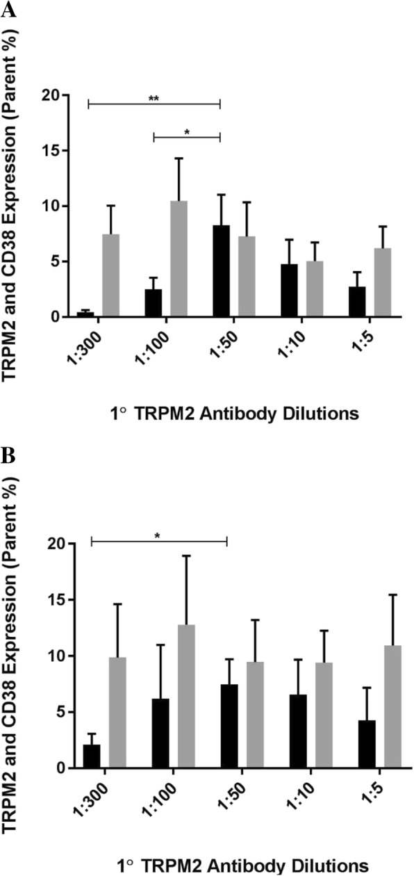
Results: At 2 h/1 h, TRPM2 (Fig. 2 A, B, p < 0.05) and TRPM2/CD38 (Fig. 3A, B, p < 0.05) surface expression significantly increased between 1:300 and 1:50 at 2 h/1 h. TRPM2/CD38 surface expression furthermore increased between 1:100 and 1:50 at 2 h/1 h (Fig. 3A, p < 0.05). Interestingly, TRPM2/CD38 surface expression significantly decreased from 1:50 to 1:5 on CD56BrightCD16Dim/- NK cells. These consistent findings highlight that 1:50 is the optimal antibody dilution and threshold to measure TRPM2 and TRPM2/CD38 surface expression on NK subsets. 2 h/1 h was determined as the optimal incubation period to ensure a sufficient timeframe for maximal antibody binding and surface expression.

Conclusion: For the first time, we describe an in vitro methodology to characterise TRPM2 and CD38 surface expression on NK cells in healthy participants. Finally, using an antibody that has not been previously applied in flow cytometry, we determined an antibody concentration and incubation time that is robust, rapid and sensitive for the application of flow cytometry.

Keywords: Antibody; Flow cytometry; Natural killer cells; Transient receptor potential Melastatin 2.


Conflict of interest statement

Ethics approval and consent to participate

All participants provided written consent and the study was approved by the Griffith University Human Research Ethics Committee (HREC/15/QGC/63).

Consent for publication

Not applicable.

Competing interests

The authors declare that they have no competing interests.

Publisher’s Note

Springer Nature remains neutral with regard to jurisdictional claims in published maps and institutional affiliations.

Figures

Fig. 1
Fig. 1
Flow cytometry gating strategy of TRPM2 and CD38 using primary TRPM2 antibody (1:50) at 2 h/1 h. a Lymphocyte populations were identified using forward and side scatter dot plots. b Duration of cell acquirement was measured followed by (c) cell viability after 7-AAD staining. d CD3+ cells were excluded and only (e) CD3- lymphocytes were further used to characterise NK cells by CD56. f CD3/CD56+ NK cells were sorted into CD56BrightCD16Dim/− and CD56Dim CD16+ NK cell subsets using CD56 and CD16. TRPM2 surface expression was measured on (g) CD56Dim CD16+ and (h) CD56BrightCD16Dim/− NK cell subsets. Dual surface expression of TRPM2 and CD38 was furthermore assessed on (i) CD56BrightCD16Dim/− and (j) CD56Dim CD16+ NK cell subsets
Fig. 2
Fig. 2
TRPM2 surface expression on Natural Killer cell subsets in healthy participants. TRPM2 surface expression was measured on (a) CD56DimCD16+ and (b) CD56BrightCD16Dim/− NK cells by flow cytometry. TRPM2 surface expression was comparatively measured at 2 h/1 h [black] and 1 h/30 min [grey] following 1:300, 1:100, 1:50, 1:10 and 1:5 primary TRPM2 antibody dilutions. Representative flow cytometry dot plots of each respective dilution are presented in Additional file 4: Figure S4, Additional file 5: Figure S5, Additional file 6: Figure S6, Additional file 7: Figure S7, Additional file 8: Figure S8, Additional file 9: Figure S9, Additional file 10: Figure S10, Additional file 11: Figure S11, Additional file 12: Figure S12, Additional file 13: Figure S13 and Additional file 17: Figure S17, Additional file 18: Figure S18, Additional file 19: Figure S19, Additional file 20: Figure S20, Additional file 21: Figure S21, Additional file 22: Figure S22, Additional file 23: Figure S23, Additional file 24: Figure S24, Additional file 25: Figure S25, Additional file 26: Figure S26. Bar graphs report the means ± SEM. Mann Whittney U test and Kruskal Wallis H test were performed. * p < 0.05. primary, TRPM2, Transient Receptor Potential Melastatin 2
Fig. 3
Fig. 3
Dual identification of TRPM2 and CD38 surface expression on Natural Killer cell subsets. TRPM2/CD38 surface expression was measured on (a) CD56DimCD16+ and (b) CD56BrightCD16Dim/− NK cells by flow cytometry. TRPM2/CD38 surface expression was comparatively measured at 2 h/1 h [black] and 1 h/30 min [grey] following 1:300, 1:100, 1:50, 1:10 and 1:5 primary TRPM2 antibody dilutions. Representative flow cytometry dot plots of each respective dilution are presented in Additional file 4: Figure S4, Additional file 5: Figure S5, Additional file 6: Figure S6, Additional file 7: Figure S7, Additional file 8: Figure S8, Additional file 9: Figure S9, Additional file 10: Figure S10, Additional file 11: Figure S11, Additional file 12: Figure S12, Additional file 13: Figure S13 and Additional file 17: Figure S17, Additional file 18: Figure S18, Additional file 19: Figure S19, Additional file 20: Figure S20, Additional file 21: Figure S21, Additional file 22: Figure S22, Additional file 23: Figure S23, Additional file 24: Figure S24, Additional file 25: Figure S25, Additional file 26: Figure S26. Bar graphs report the means ± SEM. Mann Whittney U test and Kruskal Wallis H test were performed. * p < 0.05, ** p < 0.01. 1° primary, TRPM2, Transient Receptor Potential Melastatin 2
Fig. 4
Fig. 4
Immunophenotype of TRPM2 and CD38 receptors on NK cell subsets by flow cytometry. (Source: Personal collection) a Five antibody controls were performed to determine an individualised positive TRPM2 and TRPM2/CD38 gate for each participant. Antibody controls included an unstained tube (unlabelled NK cells); secondary tube (conjugated secondary antibody FITC); and a FMO tube (CD3, CD56, CD16 and CD38). b Normal rabbit serum was used at comparable dilutions as the primary TRPM2 antibody to measure TRPM2 and TRPM2/CD38 surface expression on NK subsets. c Normalised TRPM2 and TRPM2/CD38 surface expression was calculated by compensating the percentage of fluorescence spill over into the B525_50 (TRPM2) and V525_50 (CD38) detectors from the TRPM2 antibody stained tube on both NK subsets

References

    1. Vivier E, Tomasello E, Baratin M, Walzer T, Ugolini S. Functions of natural killer cells. Nat Immunol. 2008;9(5):503. doi: 10.1038/ni1582. - DOI - PubMed
    1. Draghi M, Yawata N, Gleimer M, Yawata M, Valiante NM, Parham P. Single-cell analysis of the human NK cell response to missing self and its inhibition by HLA class I. Blood. 2005;105(5):2028–2035. doi: 10.1182/blood-2004-08-3174. - DOI - PubMed
    1. Schwarz EC, Qu B, Hoth M. Calcium, cancer and killing: the role of calcium in killing cancer cells by cytotoxic T lymphocytes and natural killer cells. Biochimic Biophys Acta. 2013;1833(7):1603–1611. doi: 10.1016/j.bbamcr.2012.11.016. - DOI - PubMed
    1. Nieto-Posadas A, Jara-Oseguera A, Rosenbaum T. TRP channel gating physiology. Curr Top Med Chem. 2011;11(17):2131–2150. doi: 10.2174/156802611796904870. - DOI - PubMed
    1. Venkatachalam K, Montell C. TRP channels. Annu Rev Biochem. 2007;76:387–417. doi: 10.1146/annurev.biochem.75.103004.142819. - DOI - PMC - PubMed

Publication types