. 2019 Jul;9(7):1249-1258.
doi: 10.1002/2211-5463.12655. Epub 2019 Jun 2.

XBP1 inhibits mesangial cell apoptosis in response to oxidative stress via the PTEN/AKT pathway in diabetic nephropathy

Affiliations

XBP1 inhibits mesangial cell apoptosis in response to oxidative stress via the PTEN/AKT pathway in diabetic nephropathy

Yan Wang et al. FEBS Open Bio. 2019 Jul.

Retraction in

Abstract

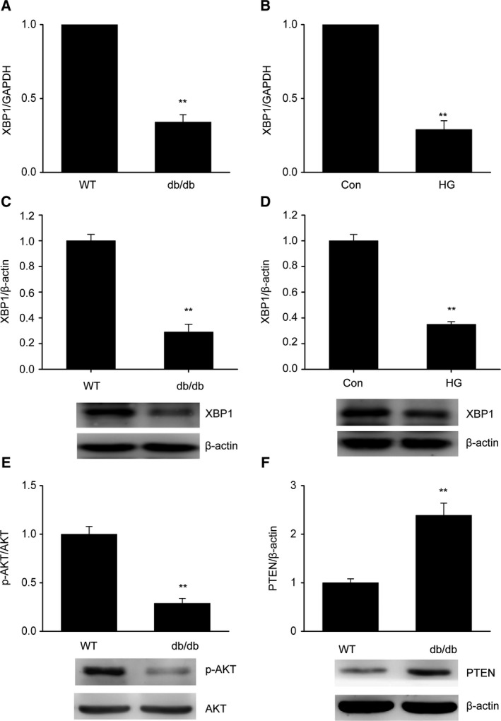
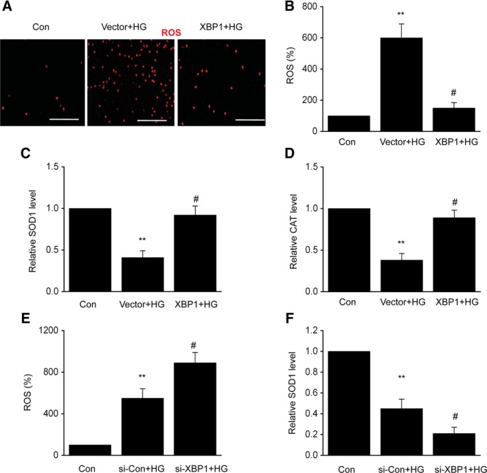
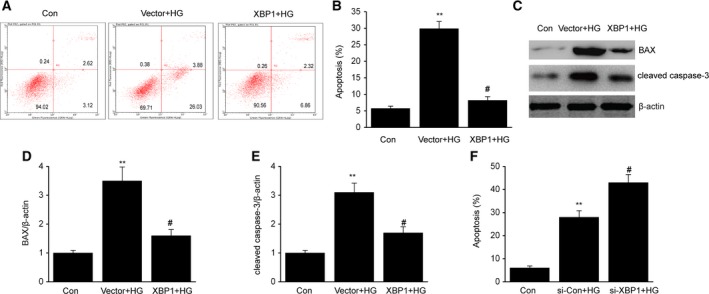
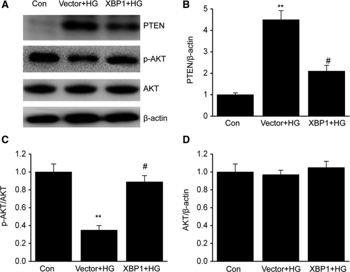
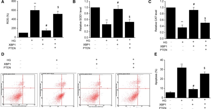
Diabetic nephropathy (DN) is a complication of diabetes mellitus (DM) that frequently results in renal disease, and is characterized by a variety of symptoms, including albuminuria. It has been shown that apoptosis of glomerular mesangial cells (MCs) can aggravate albuminuria and contribute to the development of diabetic glomerulosclerosis. Hence, determination of the mechanisms leading to MC apoptosis may help us gain insights into the pathogenesis of DN. As our understanding of the role of high glucose (HG) in MC apoptosis remains elusive, we explored the interplay between X-box binding protein 1 (XBP1) and MC apoptosis in this study. XBP1 was observed to be downregulated both in vivo and in vitro. Treatment of XBP1-overexpressing cells with HG resulted in a decrease of reactive oxygen species (ROS) and a suppression of cell apoptosis, concomitant with decreases in cleaved caspase-3 and Bax. Subsequent analyses demonstrated that XBP1 overexpression inhibited the expression of phosphatase and tensin homolog deleted on chromosome ten (PTEN) and enhanced the activation of AKT in MCs exposed to HG. In addition, XBP1-induced injuries in MC were reversed by overexpression of PTEN, and XBP1 inhibited apoptosis, which was mediated by the activated PTEN/AKT signaling pathway. Thus, our data indicate that XBP1 can activate the PTEN/AKT signaling pathway, thereby alleviating oxidative stress caused by HG or MC apoptosis. These findings suggest that XBP1 may have potential in the development of treatment methods for DN.

Keywords: PTEN/AKT pathway; XBP1; apoptosis; mesangial cell; oxidative stress.


Conflict of interest statement

The authors declare no conflict of interest.

Figures

Figure 1
Figure 1
X‐box binding protein 1 is downregulated in diabetic mouse glomeruli and in MCs exposed to HG. (A,B) Decreased expression of XBP1 in db/db mouse glomeruli (A) and in MCs treated with HG (B), as determined by quantitative reverse transcription PCR (qRT/PCR). (C,D) Decreased expression of XBP1 in db/db mouse glomeruli (C) and in MCs treated with HG (D), as determined by western blotting. (E,F) Decreased expression of p‐AKT (E) and increased expression of PTEN (F) in db/db mouse glomeruli (C) and in MCs treated with HG (D), as determined by western blotting. Data are represented as the mean ± SEM. The experiment was conducted in triplicate. **P < 0.01 vs. the control group. (unpaired t‐test).
Figure 2
Figure 2
X‐box binding protein 1 suppresses HG‐induced oxidative stress in MCs. After 24 h of transfection with an XBP1 plasmid (XBP1) or empty vector (vector), MCs were treated with HG for 48 h. (A) Intracellular generation of ROS by transforming H2DCFDA to DCF via an oxidative reaction. Scale bars: 100 μm. (B) Quantification data for the ROS level of each group. (C) SOD1 activity detection. (D) CAT activity detection. After 24 h of transfection with XBP1‐specific siRNA (si‐XBP1) or negative control (si‐Con), MCs were treated with HG for 48 h. (E) Quantification data for the ROS level of each group. (F) SOD1 activity detection. Data are represented as the mean ± SEM. The experiment was conducted in triplicate. **P < 0.01 vs. the control group; # P < 0.05 vs. the vector + HG group or the si‐Con + HG group. One‐way ANOVA with Tukey's post hoc test.
Figure 3
Figure 3
X‐box binding protein 1 attenuates HG‐induced cell apoptosis in MCs. After 24 h of transfection with an XBP1 plasmid (XBP1) or empty vector (vector), MCs were treated with HG for 48 h. (A) Representative image of cell apoptosis determined via flow cytometry. (B) Quantification data for the apoptotic cells for each group. (C–E) Representative immunoblots (C) and quantitative analysis of Bax (D) and cleaved caspase‐3 (E) in MCs. After 24 h of transfection with XBP1‐specific siRNA (si‐XBP1) or negative control (si‐Con), MCs were treated with HG for 48 h. (F) Quantification data for the apoptotic cells for each group. Data are represented as the mean ± SEM. The experiment was conducted in triplicate. **P < 0.01 vs. the control group; # P < 0.05 vs. the vector + HG group or the si‐Con + HG group. One‐way ANOVA with Tukey's post hoc test.
Figure 4
Figure 4
X‐box binding protein 1 activates the AKT signaling pathway through downregulation of PTEN in MCs. After 24 h of transfection with an XBP1 plasmid (XBP1) or empty vector (vector), MCs were treated with HG for 48 h. A–D, Representative immunoblots (A) and quantitative analysis of PTEN (B), phosphorylation of AKT (C), and total AKT (D) in MCs. Data are represented as the mean ± SEM. The experiment was conducted in triplicate. **P < 0.01 vs. the control group; # P < 0.05 vs. the vector+HG group. One‐way ANOVA with Tukey's post hoc test.
Figure 5
Figure 5
The PTEN/AKT signaling pathway is significant in mediating the protective effect of XBP1 against oxidative stress and apoptosis. After 24 h of cotransfection with an XBP1 plasmid (XBP1) or PTEN plasmid (PTEN), MCs were treated with HG for 48 h. (A) Intracellular generation of ROS by transforming H2DCFDA to DCF via an oxidative reaction. (B) SOD1 activity detection. (C) CAT activity detection. (D–E) Representative of cell apoptosis by flow cytometry. Data are represented as the mean ± SEM. The experiment was conducted in triplicate. **P < 0.01 vs. the control group; # P < 0.05 vs. the HG group; $ P < 0.05 vs. the XBP1+HG group. One‐way ANOVA with Tukey's post hoc test.

References

    1. Jiang S (2018) Precise medicine for diabetic nephropathy based on multiomics. Zhonghua Yi Xue Za Zhi 98, 1053–1056. - PubMed
    1. Vallianou N, Stratigou T, Paikopoulou A, Apostolou T, Vlassopoulou B, Tsagarakis S and Ioannidis G (2018) Monitoring of patients with type 2 diabetes and nephropathy in a specialized diabetic nephropathy clinic seems to be beneficial. Diabetes Metab Syndr 12, 689–692. - PubMed
    1. Kim YC, Shin N, Lee S, Hyuk H, Kim YH, Kim H, Park SK, Cho JH, Kim CD, Ha J et al (2018) Effect of post‐transplant glycemic control on long‐term clinical outcomes in kidney transplant recipients with diabetic nephropathy: a multicenter cohort study in Korea. PLoS ONE 13, e195566. - PMC - PubMed
    1. Abedini A and Roomizadeh P (2018) The association between apolipoprotein E polymorphism and diabetic nephropathy in Iranian patients. Saudi J Kidney Dis Transpl 29, 478–479. - PubMed
    1. Singh L, Arya A and Gupta S (2018) Role of atrial natriuretic peptide in controlling diabetic nephropathy in rats. J Basic Clin Physiol Pharmacol 29, 499–505. - PubMed

Publication types

MeSH terms