

Extraskeletal Calcifications in Hutchinson-Gilford Progeria Syndrome
- PMID: 31077852
- PMCID: PMC6628204
- DOI: 10.1016/j.bone.2019.05.008
Extraskeletal Calcifications in Hutchinson-Gilford Progeria Syndrome
Abstract
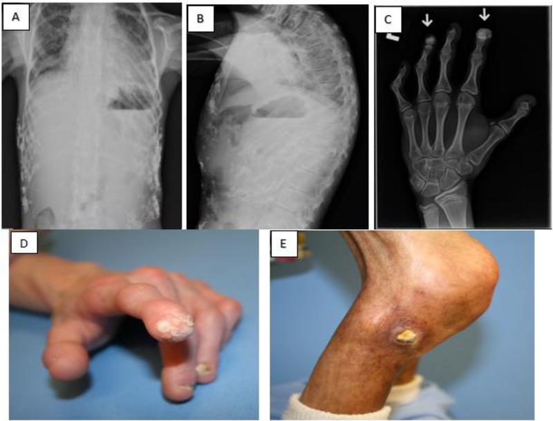
Purpose: Children with Hutchinson-Gilford progeria syndrome (HGPS), a rare premature aging disease, exhibit extraskeletal calcifications detected by radiographic analysis and on physical examination. The aim of this study was to describe the natural history and pathophysiology of these abnormal calcifications in HGPS, and to determine whether medications and/or supplements tested in clinical trials alter their development.
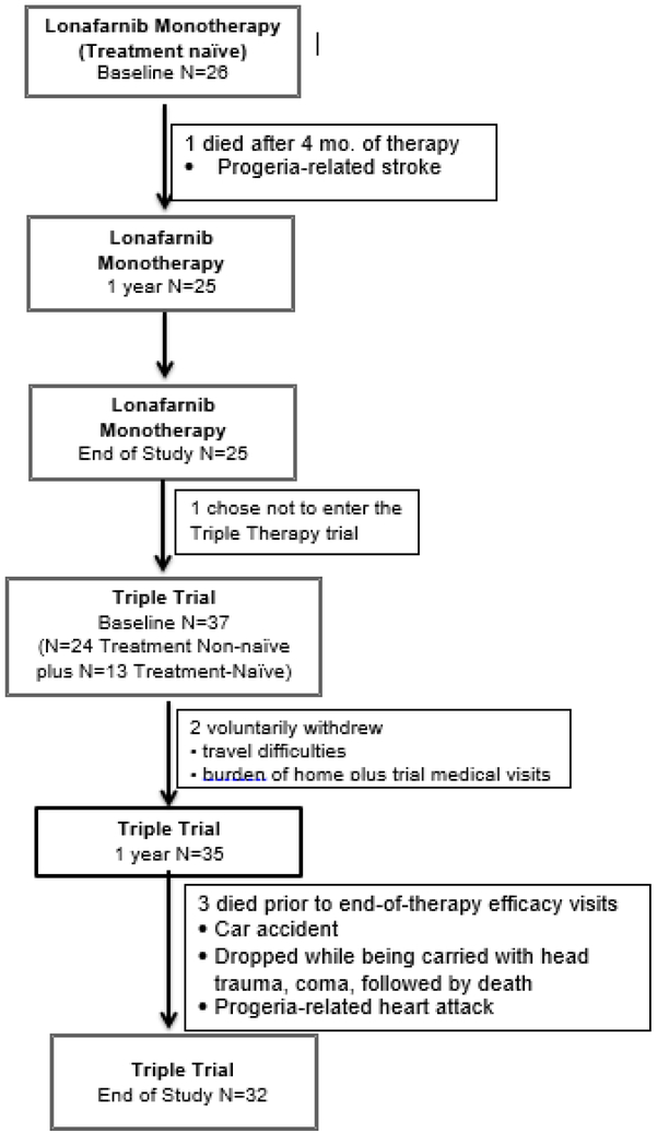
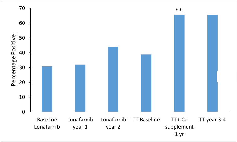
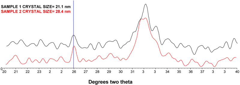
Methods: Children from two successive clinical trials administering 1) lonafarnib (n = 26) and 2) lonafarnib + pravastatin + zoledronic acid (n = 37) were studied at baseline (pre-therapy), one year on therapy, and at end-of-therapy (3.3-4.3 years after the baseline visit). Calcium supplementation (oral calcium carbonate) was administered during the first year of the second trial and was subsequently discontinued. Information on calcifications was obtained from physical examinations, radiographs, and serum and urinary biochemical measures. The mineral content of two skin-derived calcifications was determined by x-ray diffraction.
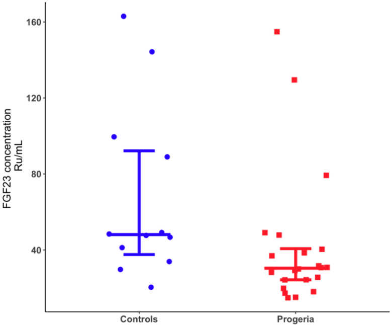
Results: Extraskeletal calcifications were detected radiographically in 12/39 (31%) patients at baseline. The odds of exhibiting calcifications increased with age (p = 0.045). The odds were unaffected by receipt of lonafarnib, pravastatin, and zoledronate therapies. However, administration of calcium carbonate supplementation, in conjunction with all three therapeutic agents, significantly increased the odds of developing calcifications (p = 0.009), with the odds plateauing after the supplement's discontinuation. Composition analysis of calcinosis cutis showed hydroxyapatite similar to bone. Although serum calcium, phosphorus, and parathyroid hormone (PTH) were within normal limits at baseline and on-therapy, PTH increased significantly after lonafarnib initiation (p < 0.001). Both the urinary calcium/creatinine ratio and tubular reabsorption of phosphate (TRP) were elevated at baseline in 22/39 (56%) and 31/37 (84%) evaluable patients, respectively, with no significant changes while on-therapy. The mean calcium × phosphorus product (Ca × Pi) was within normal limits, but plasma magnesium decreased over both clinical trials. Fibroblast growth factor 23 (FGF23) was lower compared to age-matched controls (p = 0.03).
Conclusions: Extraskeletal calcifications increased with age in children with HGPS and were composed of hydroxyapatite. The urinary calcium/creatinine ratio and TRP were elevated for age while FGF23 was decreased. Magnesium decreased and PTH increased after lonafarnib therapy which may alter the ability to mobilize calcium. These findings demonstrate that children with HGPS with normal renal function and an unremarkable Ca × Pi develop extraskeletal calcifications by an unidentified mechanism that may involve decreased plasma magnesium and FGF23. Calcium carbonate accelerated their development and is, therefore, not recommended for routine supplementation in these children.
Keywords: Aging; Extraskeletal calcifications; HGPS; Lamin; Laminopathy; Magnesium; Parathyroid hormone; Progeria.
Copyright © 2019 Elsevier Inc. All rights reserved.
Figures
References
-
- Villa-Bellosta R, Rivera-Torres J, Osorio FG, et al. Defective extracellular pyrophosphate metabolism promotes vascular calcification in a mouse model of hutchinson-gilford progeria syndrome that is ameliorated on pyrophosphate treatment. Circulation. 2013; 127: 2442–51. - PubMed
Publication types
MeSH terms
Substances
Grants and funding
LinkOut - more resources
Full Text Sources
Research Materials
Miscellaneous
