. 2019 Jul:210:70-82.
doi: 10.1016/j.biomaterials.2019.04.030. Epub 2019 May 1.

Optimizing PLG nanoparticle-peptide delivery platforms for transplantation tolerance using an allogeneic skin transplant model

Affiliations

Optimizing PLG nanoparticle-peptide delivery platforms for transplantation tolerance using an allogeneic skin transplant model

Sahil Shah et al. Biomaterials. 2019 Jul.

Abstract

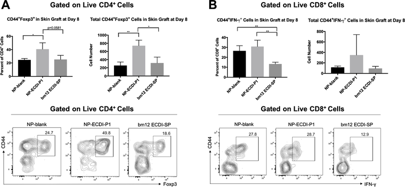
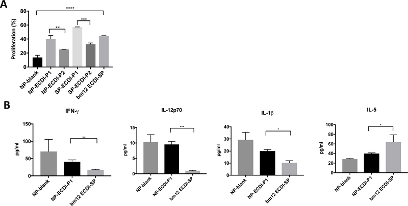
A robust regimen for inducing allogeneic transplantation tolerance involves pre-emptive recipient treatment with donor splenocytes (SP) rendered apoptotic by 1-ethyl-3-(3'-dimethylaminopropyl)-carbodiimide(ECDI) treatment. However, such a regimen is limited by availability of donor cells, cost of cell procurement, and regulatory hurdles associated with cell-based therapies. Nanoparticles (NP) delivering donor antigens are a promising alternative for promoting transplantation tolerance. Here, we used a B6.C-H-2bm12(bm12) to C57BL/6(B6) skin transplant model involving a defined major histocompatibility antigen mismatch to investigate design parameters of poly(lactide-co-glycolide) (PLG) NPs delivering peptides containing the donor antigen for optimizing skin allograft survival. We showed that an epitope-containing short peptide (P1) was more effective than a longer peptide (P2) at providing graft protection. Importantly, the NP and P1 complex (NP-ECDI-P1) resulted in a significant expansion of graft-infiltrating Tregs. Interestingly, in comparison to donor ECDI-SP that provided indefinite graft protection, NP-ECDI-P1 targeted different splenic phagocytes and skin allografts in these recipients harbored significantly more graft-infiltrating CD8+IFN-γ+ cells. Collectively, the current study provides initial engineering parameters for a cell-free and biocompatible NP-peptide platform for transplant immunoregulation. Moreover, it also provides guidance to future NP engineering endeavors to recapitulate the effects of donor ECDI-SP as a goal for maximizing tolerance efficacy of NP formulations.

Keywords: Nanoparticles; Poly(lactide-co-glycolide) (PLG); Skin transplantation; Transplantation tolerance; bm-12.


Conflict of interest statement

Conflict of Interest: The authors have no competing interests to disclose.

Figures

Figure 1.
Figure 1.. PLG NP characterization and quantification of antigen coupling.
(A) Nanoparticles size (486.5 ± 26.4 nm) and (B) surface Zeta Potential distribution (−40.95 ± 2.66 mV) were obtained using dynamic light scattering (n = 3). (C) Representative scanning Electron Micrograph (SEM) image of PLG NPs used for antigen coupling. (D) Loading amount. After peptide coupling by different methods, the amount of peptide loaded on each platform was determined by CBQCA assay. Measurements were standardized to 1 mg of PLG NPs or 3.3×10⁷ splenocytes (n = 5), as 3 mg of PLG NPs or 1×10⁸ antigen-coupled splenocytes per dose were injected for subsequent in vivo experiments. (E) Loading efficiency. Calculated as the percentage of bound peptide following the conjugation/encapsulation reaction over the input peptide at the beginning of the conjugation/encapsulation reaction. For (A) – (C), data are representative of 3 independent experiments. For (D) and (E), data shown are average from 3 independent experiments.
Figure 2.
Figure 2.. The effect of peptide length on skin allograft survival.
Indicated preparations of NPs (3mg/dose) or SPs (1×10⁸/dose) were injected (i.v.) on day −7 and +1 to mice receiving tail to back bm12 to B6 skin transplantation on day 0. Skin grafts were monitored and rejection was determined when 80% of the graft area was necrotic. Statistical significance was determined by Log-rank (Mantel-Cox) tests. ***p<0.001, ****p<0.0001.
Figure 3.
Figure 3.. Biodistribution of NP-blank, NP-ECDI-P1, or bm12 ECDI-SP in the spleen 24 hours post i.v. injection.
(A) MHC I-Ab and Rhodamine/PKH67 double positive cells in the spleen of injected mice were gated out using the spleen from un-injected mice as a negative control (left dot plots). IAb+Rhodamine/PKH67+ cells were further analyzed for CD11c, CD8α, B220 and CD11b expressions (right dot plots). Plots shown are representative of 3 mice in each group. (B) Percentages of each splenic cell population uptaking NP-blank, NP-ECDI-P1 or bm12 ECDI-SP of total IAb+Rhodamine/PKH67+ cells. Statistical significance was determined by one-way ANOVA with Sidak’s multiple comparisons test. *p<0.05, **p<0.01. Data shown were averaged from 3 mice in each group.
Figure 4.
Figure 4.. Graft-infiltrating CD4+CD44+Foxp3+ and CD8+CD44+IFN-γ+ cells in skin allografts from recipient mice treated with NP-blank, NP-ECDI-P1 or bm12 ECDI-SP.
(A) Left bar graph: Percentage of graft-infiltrating CD44+Foxp3+ cells among graft-infiltrating CD4+ cells; Right bar graph: Total number of CD4+CD44+Foxp3+ cells in skin allografts. Representative contour plots show gating for CD44+Foxp3+ cells in skin allografts from recipients of each treatment group. (B) Left bar graph: Percentage of graft-infiltrating CD44+IFN-γ+ cells among graft-infiltrating CD8+ cells; Right bar graph: Total number of CD8+ CD44+IFN-γ+ cells in skin allografts. Representative contour plots show gating for CD44+IFN-γ+ cells in skin allografts from recipients of each treatment group. For (A) and (B), skin allografts were harvested and examined on day 8 post transplantation. Statistical significance was determined by one-way ANOVA with Sidak’s multiple comparisons test. *p<0.05, **p<0.01. Data is representative or averaged from 3 mice per group.
Figure 5.
Figure 5.. In vitro bm12-specific CD4 T cell proliferation and cytokine production in response to stimulation by APCs pulsed with different bm12 antigen sources.
(A) ABM CD4+ T cell proliferation. ABM CD4+ T cells were co-cultured with B6 BMDMs pulsed with various NP and SP formulations of P1 and P2 as the source of bm12 antigen. % proliferation of ABM CD4+ T cells was determined by % of ABM CD4+ T cells with VPD450 fluorescence lower than that of undivided ABM CD4+ T cells. (B) Cytokine production from ABM CD4+ T cells co-cultured with B6 BMDMs pulsed with NP-blank, NP-ECDI-P1 or bm12 ECDI-SP. IFN-γ, IL-12p70, IL-1β, and IL-5 levels were determined by Luminex liquichip microplate reader. For both (A) and (B), statistical significance was determined by one-way ANOVA with Sidak’s multiple comparisons test or student’s t-test. *p<0.05, **p<0.01, ***p<0.001, ****p<0.0001. Data is averaged from n=2 biological replicates per group.
Figure 6.
Figure 6.. The effect of peptide conjugation chemistry on skin allograft survival.
(A) Indicated preparations of NPs (3mg/dose) were injected (i.v.) on day −7 and +1 to mice receiving tail to back bm12 to B6 skin transplantation on day 0. Skin grafts were monitored and rejection was determined as in Figure 2. Similarly, the bm12 ECDI-SP treated group served as a positive control whereas the NP-blank treated group served as a negative control. Statistical significance was determined by Log-rank (Mantel-Cox) tests. (B) ABM CD4+ T cell proliferation. ABM CD4+ T cells were co-cultured with B6 BMDMs pulsed with either NP-ECDI-P1 or NP-BMPH-P3 as the source of bm12 antigen. % proliferation of ABM CD4+ T cells was similarly determined as in Figure 5. Statistical significance was determined by one-way ANOVA with Sidak’s multiple comparisons tests. Data is averaged from n=2 biological replicates per group. For both (A) and (B), *p<0.05, ***p<0.001, ****p<0.0001, ns = no significance.
Figure 7.
Figure 7.. Surface conjugation or encapsulation of P1 peptide provides equivalent skin allograft survival.
(A) Percent of peptide release over 196 hours of NP-ECDI-P1 and NP-Encap-P1, as described in Materials and Methods. Note that the error bars are smaller than the size of symbols for the NP-Encap-P1 data points. (B) Confocal images of a single representative NP-ECDI-P1 particle and a single representative NP-Encap-P1 particle. Magnification: ×60. (C) Fluorescence intensity profiles calculated across the gray arrow in (B) of a single representative NP-ECDI-P1 particle and a single representative NP-Encap-P1 particle. (D) Indicated preparations of NPs (3mg/dose) were injected (i.v.) on day −7 and +1 to mice receiving tail to back bm12 to B6 skin transplantation on day 0. Skin graft monitoring, determination of rejection, positive and negative controls were similar to Figure 2. Statistical significance was determined by Log-rank (Mantel-Cox) tests. (E) ABM CD4+ T cell proliferation. ABM CD4+ T cells were co-cultured with B6 BMDMs pulsed with either NP-ECDI-P1 or NP-Encap-P1 as the source of bm12 antigen. % proliferation of ABM CD4+ T cells was similarly determined as in Figure 5. Statistical significance was determined by one-way ANOVA with Sidak’s multiple comparisons tests. Data is averaged from n=2 biological replicates per group. For both (D) and (E), ***p<0.001, ****p<0.0001, ns = no significance.
Figure 8.
Figure 8.. The antigenic peptide P1 provides comparable skin allograft protection as donor cell lysate.
(A) Indicated preparations of NPs (3mg/dose) or SPs (1×10⁸/dose) were injected (i.v.) on day −7 and +1 to mice receiving tail to back bm12 to B6 skin transplantation on day 0. Skin grafts were monitored and rejection was determined as in Figure 2. Statistical significance was determined by Log-rank (MantelCox) tests. (B) ABM CD4+ T cell proliferation. ABM CD4+ T cells were co-cultured with B6 BMDMs pulsed with either NP-ECDI-lysate or SP-ECDI-lysate as the source of bm12 antigen. % proliferation of ABM CD4+ T cells was similarly determined as in Figure 5. Statistical significance was determined by one-way ANOVA with Sidak’s multiple comparisons tests. Data is averaged of n=3 biological replicates per group. For (A) and (B), ns = no significance.

References

    1. Platt JL. New directions for organ transplantation. Nature. 1998;392(6679 Suppl):11–7. - PubMed
    1. Ricordi C, Strom TB. Clinical islet transplantation: advances and immunological challenges. Nat Rev Immunol. 2004;4(4):259–68. - PubMed
    1. Abecassis M, Bartlett ST, Collins AJ, Davis CL, Delmonico FL, Friedewald JJ, et al. Kidney transplantation as primary therapy for end-stage renal disease: A National Kidney Foundation/Kidney Disease Outcomes Quality Initiative (NKF/KDOQI (TM)) conference. Clin J Am Soc Nephro. 2008;3(2):471–80. - PMC - PubMed
    1. Girlanda R Deceased organ donation for transplantation: Challenges and opportunities. World J Transplant. 2016;6(3):451–9. - PMC - PubMed
    1. Duncan MD, Wilkes DS. Transplant-related immunosuppression: a review of immunosuppression and pulmonary infections. Proc Am Thorac Soc. 2005;2(5):449–55. - PMC - PubMed

Publication types

MeSH terms