Review
. 2019 Oct:60:44-52.
doi: 10.1016/j.ceb.2019.03.016. Epub 2019 May 9.

Seeing is believing: our evolving view of kinetochore structure, composition, and assembly

Affiliations
Review

Seeing is believing: our evolving view of kinetochore structure, composition, and assembly

Grace Hamilton et al. Curr Opin Cell Biol. 2019 Oct.

Abstract

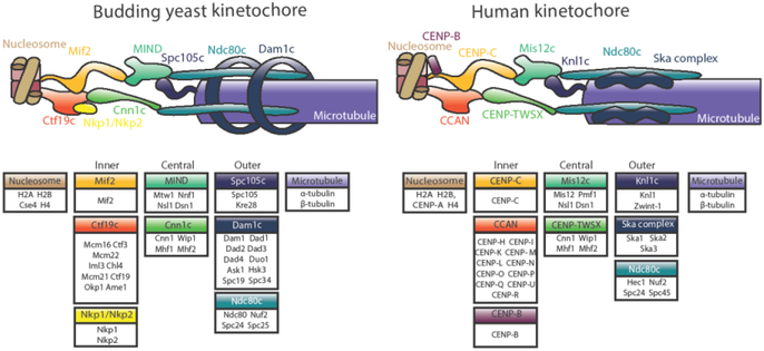
This review highlights three recent trends in the field of kinetochore biology: the proliferation of structural data for kinetochore protein complexes (including CBF3, Dam1c, Mis12cMIND, and CENP-NLChl4/Iml3); the growing consensus that the kinetochore is a dynamic structure whose composition changes as the cell cycle progresses; and the mounting evidence of multiple pathways whereby the microtubule-binding elements of the outer kinetochore may be recruited by inner kinetochore proteins. Our focus is on the two best-studied systems in the field: human and budding yeast kinetochores. This review will demonstrate the remarkable similarity of these two systems, as well as their intriguing differences.


Conflict of interest statement

Disclosure: The authors declare no conflicts of interest.

Figures

Figure 1.
Figure 1.
Model for the budding yeast (S.cerevisiae) and human (H. sapiens) kinetochores. The individual proteins of each subcomplex are shown in boxes. Inner kinetochore proteins, called the constitutively centromere-associated network (CCAN), contact the centromeric nucleosome and recruit central kinetochore proteins. These contact the microtubule-binding elements that make up the outer kinetochore. Homologous subcomplexes are colored identically, with the exception of the Dam1 and Ska complexes, which are non-homologous functional counterparts.
Figure 2.
Figure 2.
Recently published structures of outer kinetochore protein complexes. (A) Ndc80cDwarf is an elongated heterotetramer comprised of a microtubule-binding Hec1Ndc80-Nuf2 dimer and Mis12MIND-binding Spc24-Spc25 dimer. The structure shows the complete junction region of the heterotetramer (PDB ID: 5TCS) [7,8]. (B) (top) Chaetomium thermophilum Dam1c heterodecamers oligomerize into 17-membered rings with an outer diameter of about 560 Å [9]. (bottom) Ribbon diagram of a heterodecamer subunit, containing amino acids 13-78 of Ask1, 18-76 of Dad1, 25-95 and 109-116 of Dad2, 18-82 of Dad3, 3-70 of Dad4, 53-107 of Dam1, 49-121 of Duo1, 22-77 of Hsk3, 7-112 of Spc19, 3-48 and 112-199 of Spc34 (PDB ID: 6CFZ) [9]. (C) (top) K.lactis MIND is a Y-shaped heterotetramer, the stem of which binds Spc24-Spc25 of Ndc80c. One globular head binds inner kinetochore proteins CENP-CMif2 and CENP-UAme1, while a N-terminal extension of Dsn1 from the other head inhibits these interactions except when phosphorylated by Aurora B kinase (PDB ID: 5T58 and 5T51) [19]. (bottom) H.sapiens Mis12c with CENP-C shares a conserved structure with its budding yeast homolog; it also forms Y-shaped heterotetramer about 200 Å long, with conserved sites of interaction with CENP-CMif2 and Ndc80(PDB ID:5LSK) [20].
Figure 3.
Figure 3.. Recently published structures of inner kinetochore protein complexes.
(A) Dimers of the 13-protein S. cerevisiae Ctf19c flank a central cavity. CENP-NLCh14/Im13 occupies the middle of each protomer, and its CENP-ACse4-binding domain extends into the central cavity (PDB ID: 6NUW) [29]. (B) An ~22 Å negative stain 3D reconstruction of a reconstituted 11-protein human CCAN, in which CENP-HIKM (density of subcomplex in blue) and CENP-OPQUR (density of sub-complex in yellow) sandwich CENP-NL (density of subcomplex in green) [32]. (C) The N-terminus of human CENP-N specifically recognizes centromeric, CENP-A-containing nucleosomes by binding to the unique L1 loop (indicated in red) of CENP-A [27, 28]. (Model shown from PDB ID: 6C0W, related structures: 6BUZ and 6EQT) (D) Schematic of the 56 bp nuclease-resistant sequence bound to CBF3. Conserved TGT and CCG motifs in Centromere Determining Element III (CDEIII) are specifically bound by the α-MN helix and zinc finger (ZnF) motifs, respectively, of the same Cep3A subunit [32]. (E) Budding yeast CBF3 binds centromeric DNA as a dimer, recognizing specific DNA sequences (colored red) prior to Cse4 deposition [32]. Orange spheres indicate zinc atoms. (Model shown from PDB ID: 6GYS, similar structures: 6GSA and 6F07).

References

    1. Cheeseman IM, Desai A: Molecular architecture of the kinetochore-microtubule interface. Nat Rev Mol Cell Biol 2008, 9:33–46. - PubMed
    1. Nicklas RB, Ward SC: Elements of error correction in mitosis: microtubule capture, release, and tension. J Cell Biol 1994, 126:1241–1253. - PMC - PubMed
    1. Akiyoshi B, Sarangapani KK, Powers AF, Nelson CR, Reichow SL, Arellano-Santoyo H, Gonen T, Ranish JA, Asbury CL, Biggins S: Tension directly stabilizes reconstituted kinetochore-microtubule attachments. Nature 2010, 468:576. - PMC - PubMed
    1. Musacchio A, Desai A: A Molecular View of Kinetochore Assembly and Function. Biology (Basel) 2017, 6. - PMC - PubMed
    1. Bai XC, McMullan G, Scheres SHW: How cryo-EM is revolutionizing structural biology. Trends in Biochemical Sciences 2015, 40:49–57. - PubMed

Publication types

Substances