Review
. 2019 Sep:211:64-83.
doi: 10.1016/j.trsl.2019.04.004. Epub 2019 Apr 20.

3D Bioprinting of cardiac tissue and cardiac stem cell therapy

Affiliations
Review

3D Bioprinting of cardiac tissue and cardiac stem cell therapy

Matthew Alonzo et al. Transl Res. 2019 Sep.

Abstract

Cardiovascular tissue engineering endeavors to repair or regenerate damaged or ineffective blood vessels, heart valves, and cardiac muscle. Current strategies that aim to accomplish such a feat include the differentiation of multipotent or pluripotent stem cells on appropriately designed biomaterial scaffolds that promote the development of mature and functional cardiac tissue. The advent of additive manufacturing 3D bioprinting technology further advances the field by allowing heterogenous cell types, biomaterials, and signaling factors to be deposited in precisely organized geometries similar to those found in their native counterparts. Bioprinting techniques to fabricate cardiac tissue in vitro include extrusion, inkjet, laser-assisted, and stereolithography with bioinks that are either synthetic or naturally-derived. The article further discusses the current practices for postfabrication conditioning of 3D engineered constructs for effective tissue development and stability, then concludes with prospective points of interest for engineering cardiac tissues in vitro. Cardiovascular three-dimensional bioprinting has the potential to be translated into the clinical setting and can further serve to model and understand biological principles that are at the root of cardiovascular disease in the laboratory.


Figures

Figure 1:
Figure 1:
Cells for cardiac tissue engineering. These cells can be derived from multiple stem cell sources as shown in the figure. Reproduced with permission from [15].
Figure 2:
Figure 2:
(a) Steps involved in the prefabrication of 3D printed patient-specific models [31]. (b) a 3D printed luminal replica of an aberrant retro-esophageal left subclavian artery and right aortic arch for intraoperative use as congenital defect model [39].
Figure 3:
Figure 3:
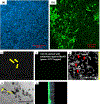
(a) illustrates the viability of mouse MSCs stained with Hoechst (blue), 24 hr after printing (b) shows the retention of mouse MSC, pre-stained with PKH67 (green), within the bioprinted construct after 5 days of culture. In (c) and (d), shown are bright field z-scans of STO fibroblasts (elongated spindle shaped) co-cultured with C2C12 myoblasts cells [rounded enlarged, confirmed in (e) and in (g)]. Scale bar is 150 μm in (c), (d) and 200 μm in (e) and (g). In (e), a single plane (cross section) was imaged whereas in (g), a Z-scan was run spanning several planes as indicated with the arrow (right hand side). In (f), a single slice of z-stack section showing top layer (fluorescent: green for C2C12) and bottom layer (non-fluorescent: STO) is shown. Reproduced with permission from [19].
Figure 4:
Figure 4:
Human vasculature printed using a modified thermal inkjet printer. Samples indicating different positions of the printed grid pattern showing the proliferation of HVECs after (a) 24 hr of culture (b and c) 7 days of culture and (d) 21 days of culture. The cells were able to network and proliferate to form a confluent lining.(e) shows the fluorescent stained printed cells that are aligned within the fibrin scaffold (f) DIC image of the fibrin scaffold (g) Confocal series of images showing the proliferated HMVEC sealed inside of the fibrin channel and the formation of a distinct tubular structure. Reproduced with permission from [61].
Figure 5.
Figure 5.
Bioprinting using Laser Induced Forward Transfer (LIFT). (a) LIFT diagram; (b) HUVECs were printed in a grid arrangement with hMSCs being printed in a square arrangement within the HUVEC grid lines. Reproduced with permission from [75].
Figure 6:
Figure 6:
Overview of the design and fabrication of structures by stereolithography (SLA). Computer-aided design (CAD) is used to produce 3D models with desired architectures. Designed models are then sliced into a series of 2D images, which are transferred to the SLA device. By sequential photo- crosslinking, 3D scaffolds are built which are exact replicas of designed 3D models. Reproduction with permission from [84].
Figure 7:
Figure 7:
Fibrin and myofibroblasts cultured in different aprotinin concentration to control the degradation of fibrin gel. (A) model of a valve conduit based on a molded fibrin gel scaffold [105]; (B) fibrin gel cultured in lower concentration of aprotinin (5 mg), fibrin gel degraded and the structure appeared to be a thin cellular layer [109]; (C) fibrin gel cultured in high concentration of aprotinin (20 mg/ml), the cell-fibrin gel structure demonstrated a multilayer-structure with myofibroblasts surrounded by extracellular matrix [109].
Figure 8:
Figure 8:
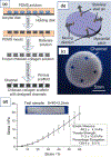
Formation and mechanical stimulation of chitosan-collagen scaffold. (a) Scheme for chitosan-collagen scaffold formation; (b) Scheme depicting mechanical stimulation applied to chitosan-collagen scaffold; (c) Chitosan-collagen scaffold; (d) Stress/strain graph. Reproduced with permission from [120].

References

    1. Organization, W.H., Global Health Estimates 2016: Deaths by Cause, Age, Sex, by Country and by Region. 2018: Geneva.
    1. Duan B, State-of-the-Art Review of 3D Bioprinting for Cardiovascular Tissue Engineering. Annals of Biomedical Engineering, 2017. 45(1): p. 195–209. - PubMed
    1. Mario Gaudino UB, Stephen Fremes, et al., Radial-Artery or Saphenous-Vein Grafts in Coronary-Artery Bypass Surgery. The New England Journal of Medicine, 2018. 378(22): p. 2069–2077. - PubMed
    1. Chadi Alraies M, P.E., Adult heart transplant: indications and outcomes. Journal of Thoracic Disease, 2014. 6(8): p. 1120–1128. - PMC - PubMed
    1. Manji Rizwan A., Burcin Ekser AHM, et al., Porcine bioprosthetic heart valves: The next generation. American Heart Journal, 2012. 164(2): p. 177–185. - PubMed

Publication types